Copyright
©The Author(s) 2017.
World J Cardiol. Jul 26, 2017; 9(7): 634-639
Published online Jul 26, 2017. doi: 10.4330/wjc.v9.i7.634
Published online Jul 26, 2017. doi: 10.4330/wjc.v9.i7.634
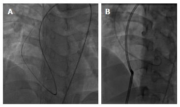
Figure 1 Through the defect, the Terumo wire was introduced to the right atrial and superior vena cava from left ventrical (A) and the 8F delivery catheter went from inferior vena cava and right atrial to the left atrial and aorta (B).
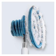
Figure 2 The Nit-Occlud® Lê VSD coil.
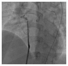
Figure 3 The 12 mm × 6 mm Nit-Occlud® Lê VSD coil was deployed with aortic approach.
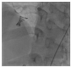
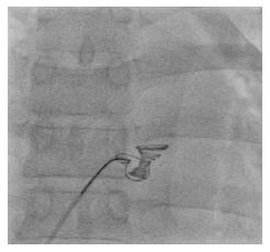
Figure 4 The 12 mm × 6 mm Nit-Occlud® Lê VSD coil immediately after release.
Figure 5 The drifting coil was retrieved with a multi-snare from the atrial side.
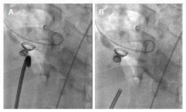
Figure 6 The bigger 14 mm × 8 mm Nit-Occlud® Lê VSD coil before (A) and immediately after release (B).
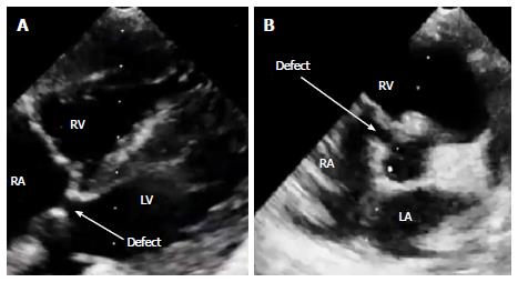
Figure 7 The Gerbode defect were best seen on echocardiography at subcostal five chamber view (A) or parasternal short axis view (B).
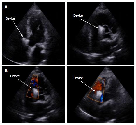
Figure 8 Six months following up showed steady result with good device position and no shunt on echocardiography.
A: Transthoracic echocardiography performed 6 mo after the procedure showed good device position at apical five chamber view (left) and parasternal short axis view (right); B: Colour doppler showed no residual shunt at apical five chamber view (left) and parasternal short axis view (right) 6 mo after device deployment.
- Citation: Phan QT, Kim SW, Nguyen HL. Percutaneous closure of congenital Gerbode defect using Nit-Occlud® Lê VSD coil. World J Cardiol 2017; 9(7): 634-639
- URL: https://www.wjgnet.com/1949-8462/full/v9/i7/634.htm
- DOI: https://dx.doi.org/10.4330/wjc.v9.i7.634


