Copyright
©The Author(s) 2017.
World J Cardiol. Jun 26, 2017; 9(6): 470-480
Published online Jun 26, 2017. doi: 10.4330/wjc.v9.i6.470
Published online Jun 26, 2017. doi: 10.4330/wjc.v9.i6.470
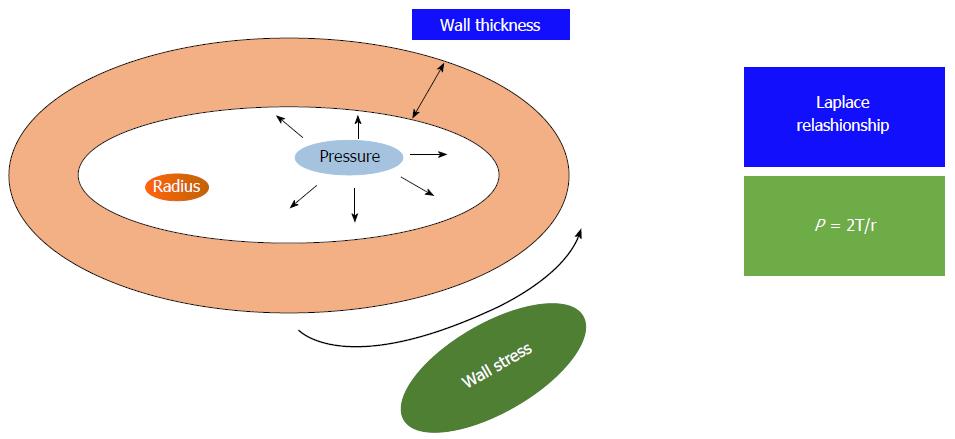
Figure 1 The figure shows the Laplace relationship: The pressure (P) generated in a sphere is directly proportional to the wall tension (T) and inversely related to the radius of the sphere (r).
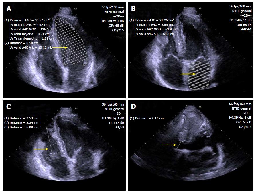
Figure 2 Standard B-mode echocardiography of endurance athlete showing enlargement of left ventricular (A), left atrial (B) and right ventricular (C) chambers, as well as inferior cavae vein dilatation (D) (arrows).
LV: Left ventricular.
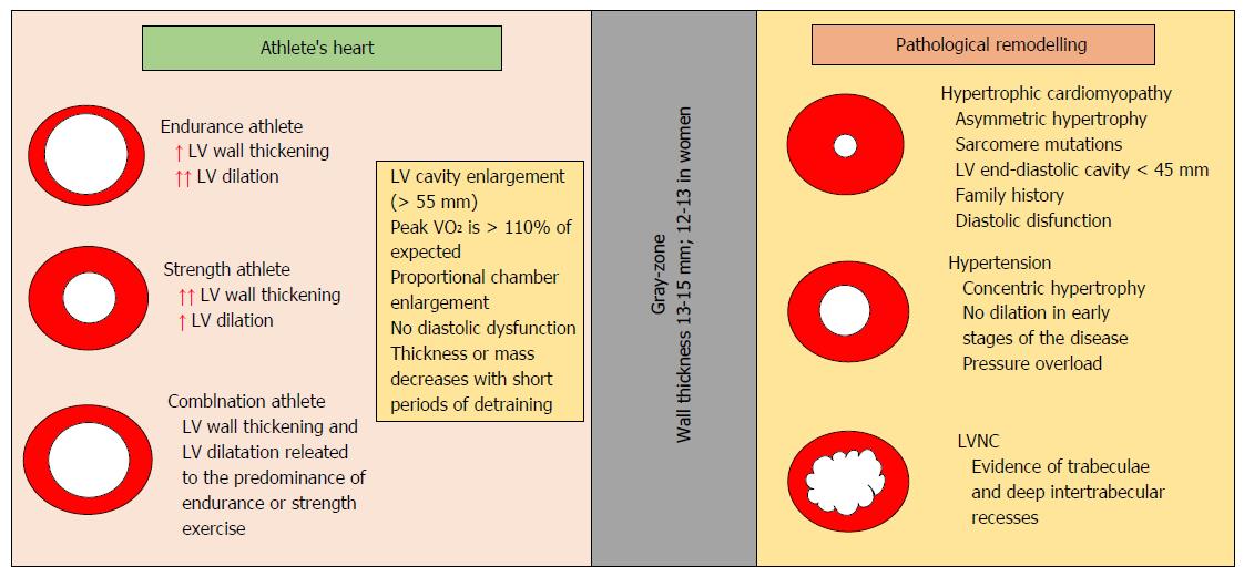
Figure 3 Different characteristics of physiological remodeling and pathological condition of the left ventricle.
LV: Left ventricular; LVNC: LV noncompaction.
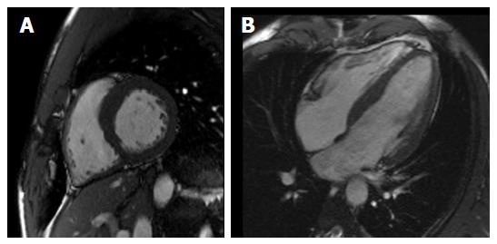
Figure 4 Cardiac magnetic resonance depicting in short-axis (A) and long-axis (B) view balanced biventricular enlargement in endurance athlete.
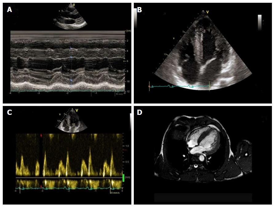
Figure 5 Non-invasive evaluation of power athlete abusing of steroids.
Standard M-mode (A) and 4-chamber view B-mode (B) echocardiography, evidencing sever left ventricular hypertrophy, with diastolic dysfunction (C) underlined by Doppler transmitral flow pattern. Cardiac magnetic resonance confirmed severe left ventricular hypertrophy (D).
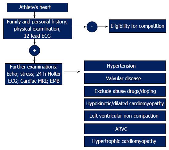
Figure 6 The management of athlete’s heart.
The figure shows an algorithm to distinguish athlete’s heart from pathological conditions. ARVC: Arrhythmogenic right ventricular cardiomyopathy; ECG: Electrocardiograph; MRI: Magnetic resonance imaging.
- Citation: Carbone A, D’Andrea A, Riegler L, Scarafile R, Pezzullo E, Martone F, America R, Liccardo B, Galderisi M, Bossone E, Calabrò R. Cardiac damage in athlete’s heart: When the “supernormal” heart fails! World J Cardiol 2017; 9(6): 470-480
- URL: https://www.wjgnet.com/1949-8462/full/v9/i6/470.htm
- DOI: https://dx.doi.org/10.4330/wjc.v9.i6.470


