Copyright
©The Author(s) 2016.
World J Cardiol. Jun 26, 2016; 8(6): 379-382
Published online Jun 26, 2016. doi: 10.4330/wjc.v8.i6.379
Published online Jun 26, 2016. doi: 10.4330/wjc.v8.i6.379
Figure 1 Coronary computed tomography shows coronary anomaly; right coronary artery from left coronary sinus running between aorta and pulmonary trunk causing functional stenosis of proximal segment (white arrow).
A: Diastole state; B: Systole state. The coronary artery at diastole state is more occlusion. Coronary angiography (C, white line arrow) shows right coronary artery originated from the left sinus of valsalva and suspicious significant stenosis of right coronary artery ostium.
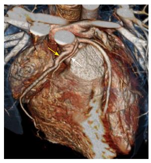
Figure 2 Coronary computed tomography after neo-ostium formation of right coronary artery with saphenous vein graft operation.
Yellow arrow is the graft vessel and white arrow is the original right coronary artery.
- Citation: Park JW, Lee JH, Kim KS, Bang DW, Hyon MS, Lee MH, Park BW. Successful extracorporeal life support in sudden cardiac arrest due to coronary anomaly. World J Cardiol 2016; 8(6): 379-382
- URL: https://www.wjgnet.com/1949-8462/full/v8/i6/379.htm
- DOI: https://dx.doi.org/10.4330/wjc.v8.i6.379


