Copyright
©The Author(s) 2015.
World J Cardiol. Oct 26, 2015; 7(10): 583-593
Published online Oct 26, 2015. doi: 10.4330/wjc.v7.i10.583
Published online Oct 26, 2015. doi: 10.4330/wjc.v7.i10.583
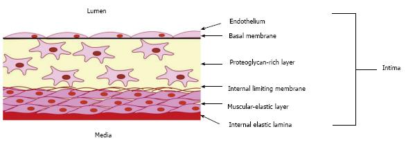
Figure 1 Schema showing the organization of the intima of the arterial wall.
The proteoglycan-rich layer which contains a heterogeneous population of cells, including macrovascular pericytes, is located just below the endothelial monolayer. Intimal pericytes form a network of cells interconnected through gap junctions. The muscular-elastic layer, formed by elongated contractile smooth muscular cells, lies below the proteoglycan-rich layer.
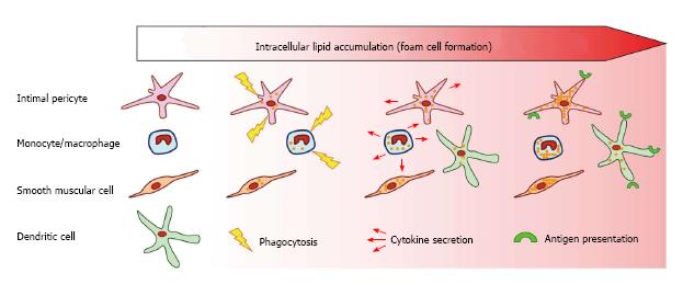
Figure 2 Scheme showing the roles of arterial wall cells in atherogenesis.
Several types of arterial wall cells participate in lipid accumulation and foam cell formation. Macrophages and intimal pericytes accumulate lipids through phagocytosis and participate in the local inflammatory process secreting pro-inflammatory cytokines. Dendritic cells, together with macrophages and intimal pericytes express antigen-presenting complexes, further promoting the inflammatory process.
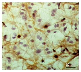
Figure 3 Cellular network formed by 3G5+ cells located under the luminal endothelium in undiseased intima.
This cellular network was identified by means of the application of peroxidase-anti-peroxidase immunohistochemical analysis (brown colour of reaction product) in en face preparation of a tissue specimen of the human aorta. Counterstain with haematoxylin.
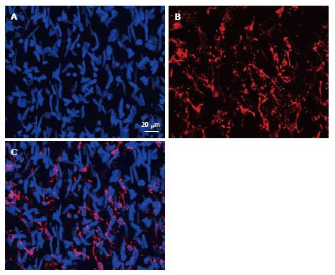
Figure 4 Immunofluorescent images representing the merge of 45 optic “confocal” sections of the intima obtained with interval of 1 μm between consecutive sections (A and B).
A: Nuclei were visualized by staining with ethidium bromide; B: Distribution and networks formed by HLA-DR+ cells, detected with the use of anti-HLA-DR antibody conjugated with Alexa 633; C: A merge of the images shown in (A) and (B). Human aorta specimen. Reproduced from Bobryshev et al[74], with permission from Elsevier. HLA: Human leukocyte antigen.
- Citation: Ivanova EA, Bobryshev YV, Orekhov AN. Intimal pericytes as the second line of immune defence in atherosclerosis. World J Cardiol 2015; 7(10): 583-593
- URL: https://www.wjgnet.com/1949-8462/full/v7/i10/583.htm
- DOI: https://dx.doi.org/10.4330/wjc.v7.i10.583


