Copyright
©2014 Baishideng Publishing Group Inc.
World J Cardiol. Sep 26, 2014; 6(9): 1041-1044
Published online Sep 26, 2014. doi: 10.4330/wjc.v6.i9.1041
Published online Sep 26, 2014. doi: 10.4330/wjc.v6.i9.1041
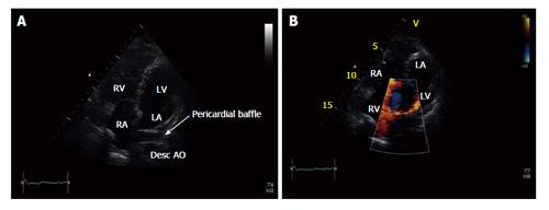
Figure 1 Apical 4-chamber view.
A: The Mustard procedure employs a pericardial baffle to direct systemic venous return to the left ventricle and pulmonary venous return to the right ventricle; B: With color Doppler. The deoxygenated blood from the vena cavae is directed to the mitral valve and and thence into the left ventricle which is the pumping ventricle for the pulmonary artery and the pulmonary circulation. LV: Left ventricle; RV: Right ventricle.
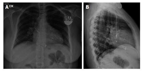
Figure 2 Chest X-ray.
A: Antero-posterior chest X-ray after pacemaker implantation to confirm the position of the pacemaker leads. The ventricular lead is situated in the anatomic left ventricle, and the atrial lead in the systemic venous baffle; B: Lateral chest X-ray after pacemaker implantation to confirm the position of the pacemaker leads In this patient the left atrial appendage was kept outside of venous tissue therefore the atrial lead was inserted and screwed into the systemic venous channel and a loop was created.
- Citation: Puntrello C, Lucà F, Rubino G, Rao CM, Gelsomino S. Systemic venous atrium stimulation in transvenous pacing after mustard procedure. World J Cardiol 2014; 6(9): 1041-1044
- URL: https://www.wjgnet.com/1949-8462/full/v6/i9/1041.htm
- DOI: https://dx.doi.org/10.4330/wjc.v6.i9.1041


