Copyright
©2012 Baishideng Publishing Group Co.
World J Cardiol. Apr 26, 2012; 4(4): 121-127
Published online Apr 26, 2012. doi: 10.4330/wjc.v4.i4.121
Published online Apr 26, 2012. doi: 10.4330/wjc.v4.i4.121
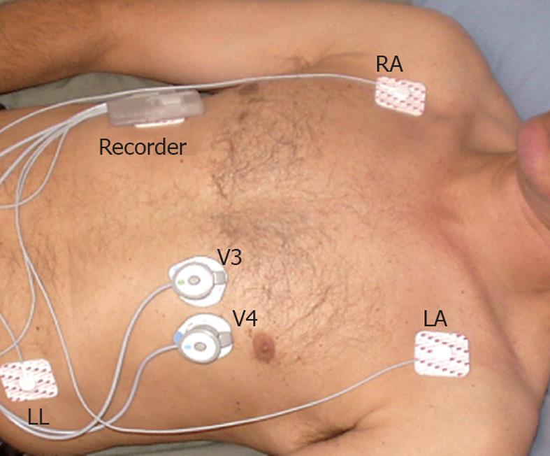
Figure 1 Placement of the recorder unit and the electrocardiography /sound sensors.
Regular electrocardiography (ECG) monitoring electrodes are placed on the right arm (RA), left arm (LA) and left leg (LL) locations for ECG monitoring. Combined ECG/sound sensors are placed in the precordial V3 and V4 positions. The recorder unit is placed on the right upper chest wall.
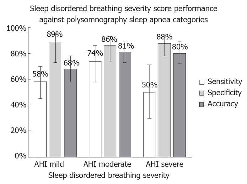
Figure 2 Performance of sleep-disordered breathing severity score against polysomnography apnea/hypopnea index for categorizing subjects into mild, moderate or severe sleep apnea on test set data.
Error bars show 95% confidence intervals. AHI: Apnea/hypopnea index.
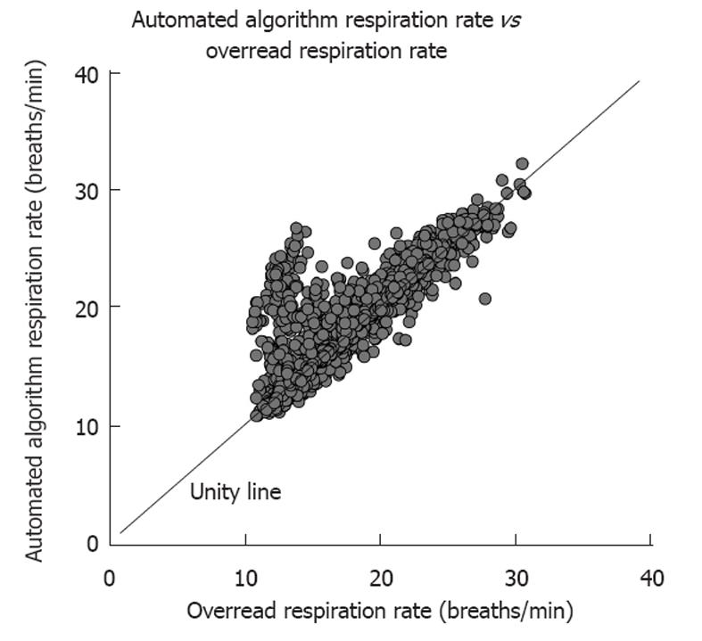
Figure 3 Relationship of the automated respiration rate algorithm to the over-read respiration rate on test set data.
- Citation: Dillier R, Baumann M, Young M, Erne S, Schwizer B, Zuber M, Erne P. Continuous respiratory monitoring for sleep apnea screening by ambulatory hemodynamic monitor. World J Cardiol 2012; 4(4): 121-127
- URL: https://www.wjgnet.com/1949-8462/full/v4/i4/121.htm
- DOI: https://dx.doi.org/10.4330/wjc.v4.i4.121


