Copyright
©2010 Baishideng Publishing Group Co.
World J Cardiol. Dec 26, 2010; 2(12): 437-442
Published online Dec 26, 2010. doi: 10.4330/wjc.v2.i12.437
Published online Dec 26, 2010. doi: 10.4330/wjc.v2.i12.437
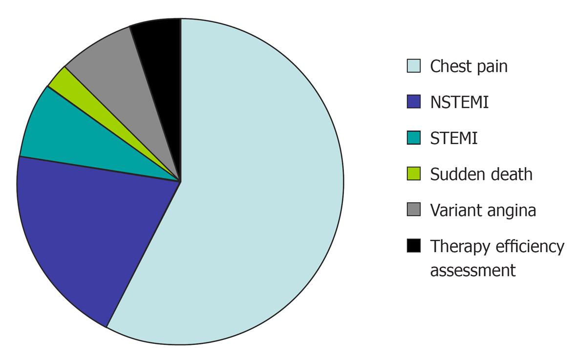
Figure 1 Indications for ergonovine stress echocardiography (n = 40).
In all cases with myocardial infarction and referred for ergonovine stress echocardiography, coronary angiography did not show significant stenosis, and a vasospasm provocation technique was performed to orientate the study of its aetiology. Ergonovine echocardiography for studying chest pain was performed when there was a suspicion of coronary vasospasm due to clinical characteristics. We defined variant angina when there was documentation of transient ST-segment elevation in patients with chest pain with normal blood levels of necrosis biomarkers. NSTEMI: Non ST elevation myocardial infarction; STEMI: ST elevation myocardial infarction.
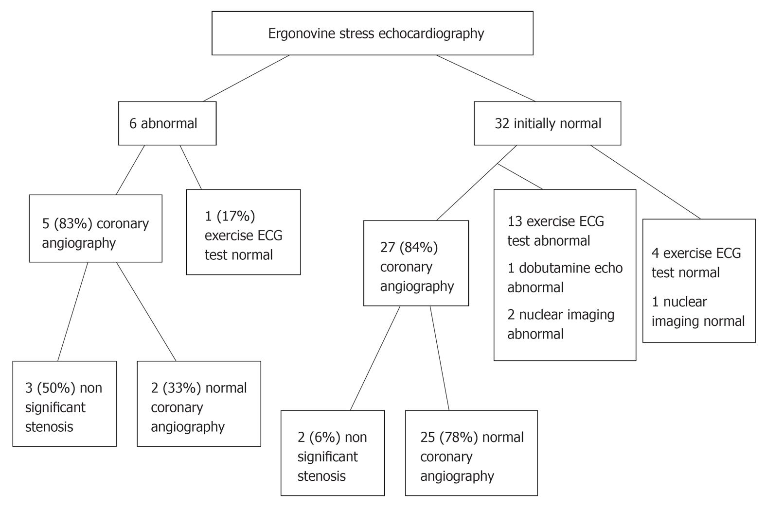
Figure 2 Tests performed before ergonovine stress echocardiography depending on the results of the stress test.
ECG: Electrocardiography.
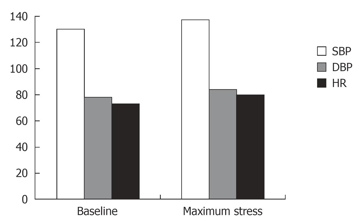
Figure 3 Blood pressure and heart rate at baseline and during the maximum stress stage of ergonovine stress echocardiography.
DBP: Diastolic blood pressure (mmHg); HR: Heart rate (beats/min); SBP: Systolic blood pressure (mmHg).
- Citation: Cortell A, Marcos-Alberca P, Almería C, Rodrigo JL, Pérez-Isla L, Macaya C, Zamorano JL. Ergonovine stress echocardiography: Recent experience and safety in our centre. World J Cardiol 2010; 2(12): 437-442
- URL: https://www.wjgnet.com/1949-8462/full/v2/i12/437.htm
- DOI: https://dx.doi.org/10.4330/wjc.v2.i12.437


