Copyright
©The Author(s) 2025.
World J Cardiol. May 26, 2025; 17(5): 105670
Published online May 26, 2025. doi: 10.4330/wjc.v17.i5.105670
Published online May 26, 2025. doi: 10.4330/wjc.v17.i5.105670
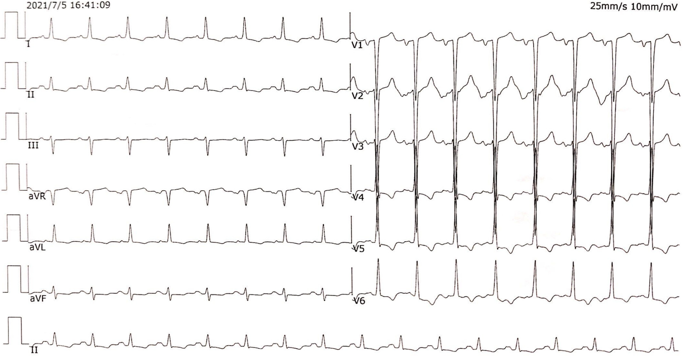
Figure 1 Electrocardiogram showing sinus tachycardia with high voltage in the left ventricle and diffuse ST-T changes.
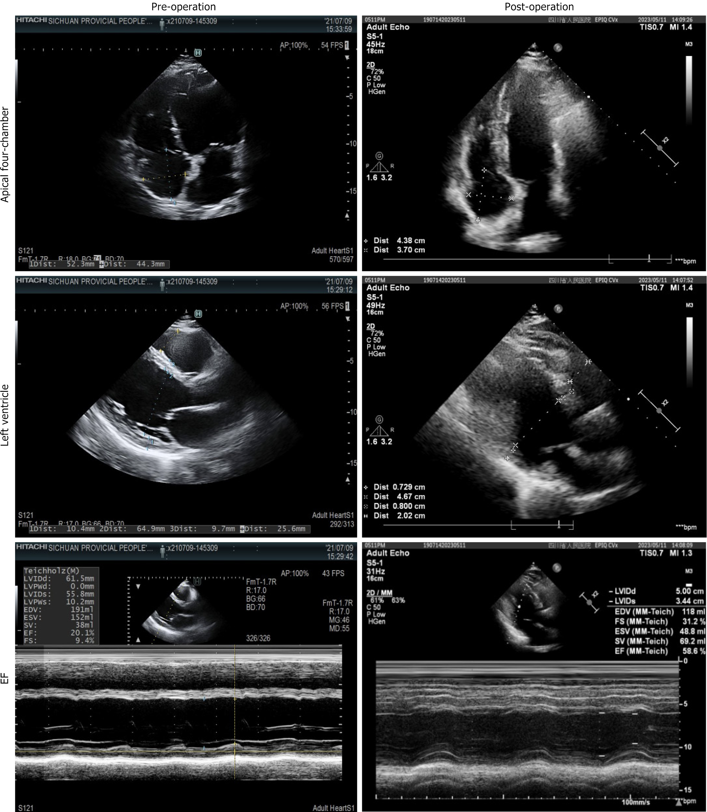
Figure 2 Transthoracic echocardiogram of preoperation and postoperation obtained in July 2021 and May 2023 respectively.
Parasternal long-axis (M-mode) and apical four-chamber views show significant changes in left ventricle, left atrium, and left ventricular ejection fraction from preoperation to postoperation. EF: Ejection fraction.
Figure 3 Findings on parathyroid ultrasonography.
A: A hypoechoic, well-circumscribed mass located posteriorly inferiorly to the lower pole of the right thyroid lobe; B: Color doppler scan showing increased vascular flow.
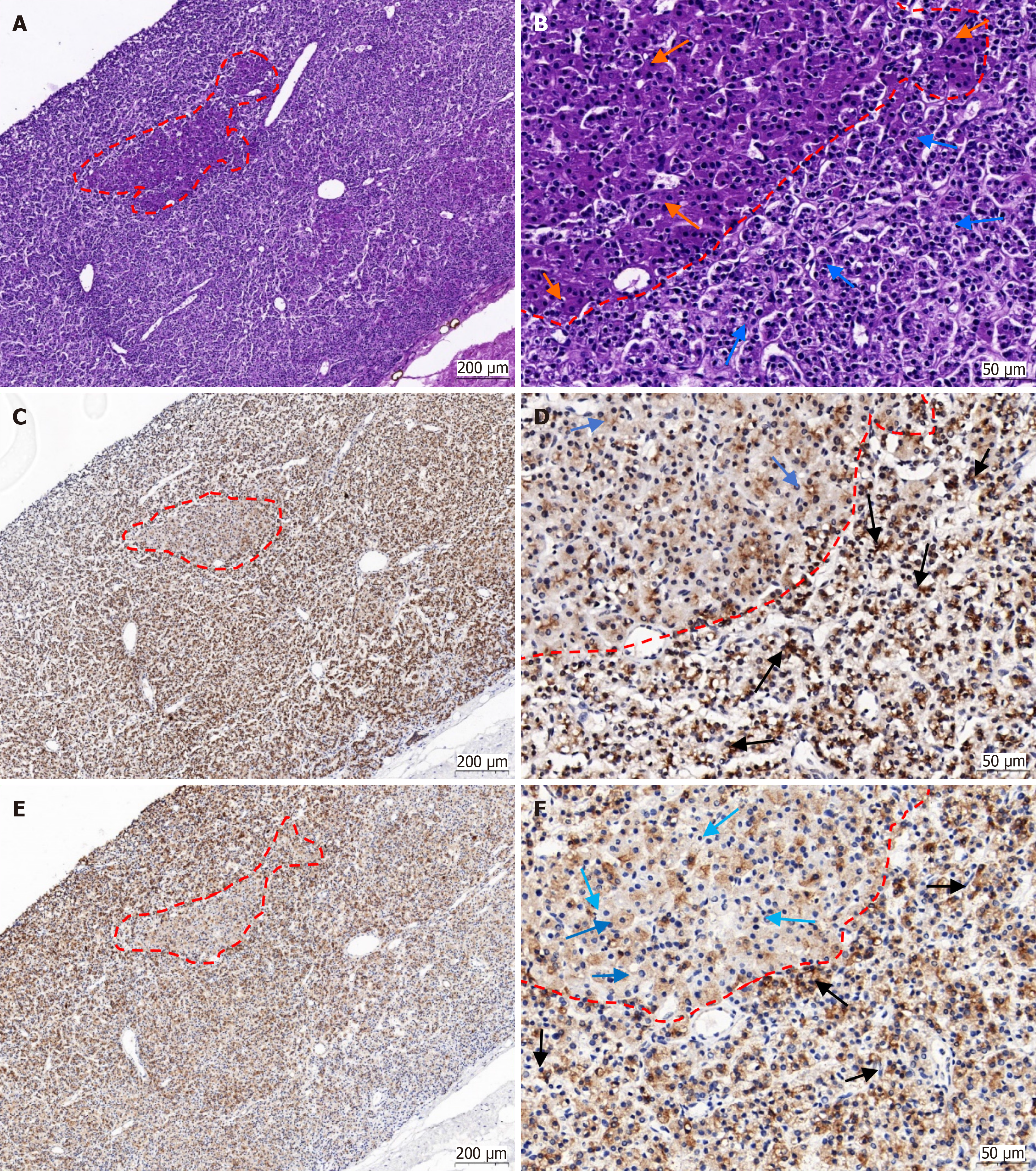
Figure 4 Histopathology of the parathyroid adenoma in this patient.
A and B: Original magnification 10.0 × and 40 ×. In the tissue of parathyroid adenoma, the structures of main cells and eosinophils are clearly distinguishable on hematoxylin and eosin stained sections, and eosinophils aggregate into clusters, significantly increasing compared to normal parathyroid glands. Orange: Eosinophils, blue: Main cell. The dashed line indicates the boundary between eosinophil clusters and main cells; C and D: Positive precipitates of parathyroid hormone immunohistochemistry (IHC) staining can be seen in the cytoplasm of main cells and eosinophils. Among them, the cytoplasmic positivity intensity of the main cells is high, eosinophil positive particles are mainly distributed at the top of the cells, and negative around the nucleus. Black: Positive staining of main cells, blue: Positive staining of eosinophils. The dashed line indicates the boundary between eosinophil clusters and main cells; E and F: The strong positive result of synaptophysin IHC can be seen in the cytoplasm of the main cells. Some eosinophils are weakly positive, while others are negative. Black: Positive staining of main cells, blue: Positive staining of eosinophils, light blue: Negative eosinophils. The dashed line indicates the boundary between eosinophil clusters and main cells.
Figure 5 Under emission computed tomography scanning, the color of the parathyroid adenoma is darker and darker than that of the thyroid gland.
The green dashed circle indicates the thyroid tissue, and the blue dashed circle indicates the parathyroid adenoma tissue.
- Citation: Jiang W, Qiu YZ, Xi HT, Ma HH, Wu X, Yuan XM, Wang WY, Kong H, Li XP. Reversible dilated cardiomyopathy caused by primary hyperparathyroidism: A case report. World J Cardiol 2025; 17(5): 105670
- URL: https://www.wjgnet.com/1949-8462/full/v17/i5/105670.htm
- DOI: https://dx.doi.org/10.4330/wjc.v17.i5.105670


