Copyright
©The Author(s) 2021.
World J Cardiol. Jun 26, 2021; 13(6): 177-182
Published online Jun 26, 2021. doi: 10.4330/wjc.v13.i6.177
Published online Jun 26, 2021. doi: 10.4330/wjc.v13.i6.177
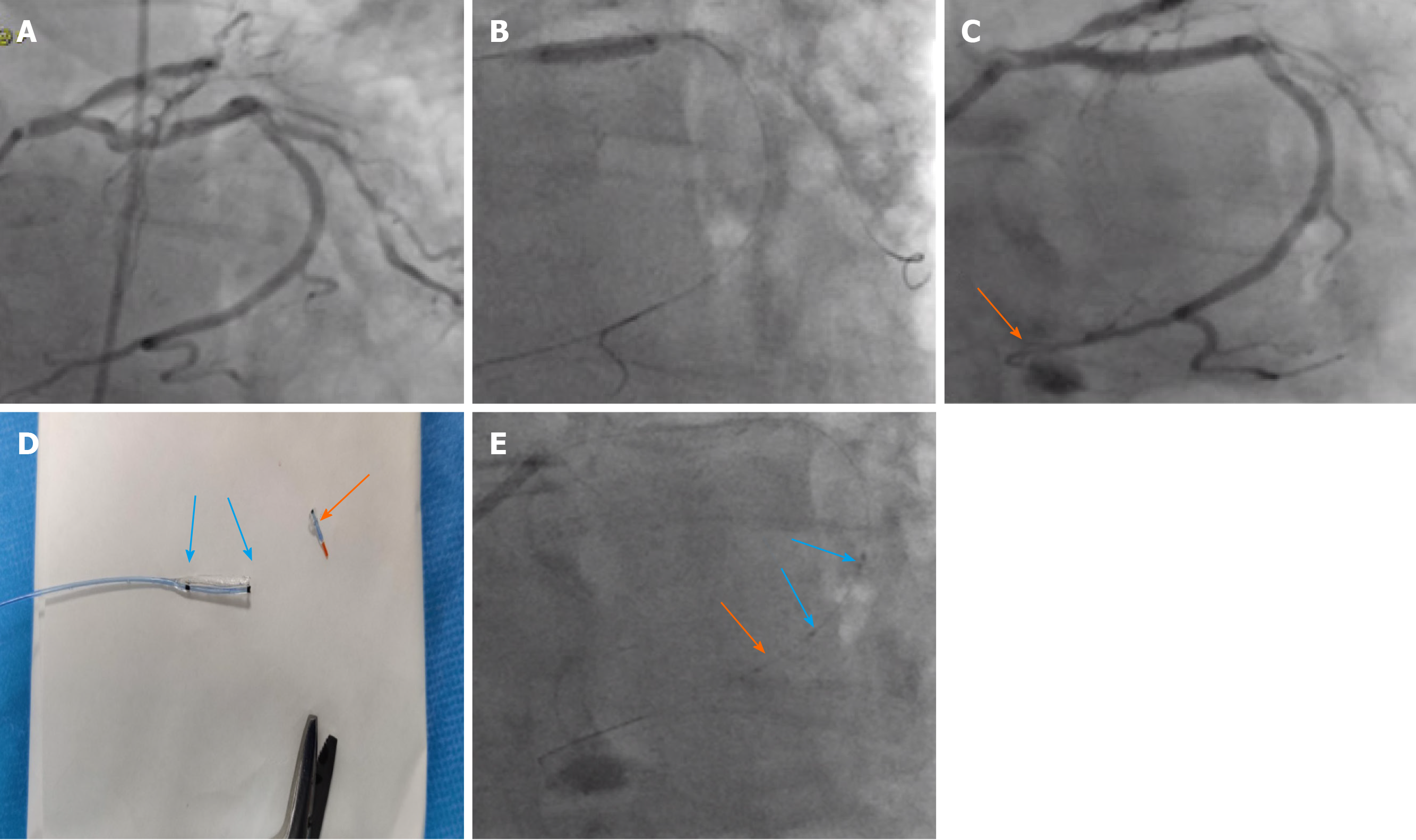
Figure 1 The proximal left circumflex artery.
A: Calcific proximal dominant left circumflex artery (LCX) lesion; B: Distal wire position when predilatation; C: Distal wire perforation in the LCX (orange arrow); D: Tip of the balloon cut for embolization; E: The cut balloon tip with the marker for X-Ray visibility (orange arrow) pushed by another intact balloon with two markers (blue arrows).
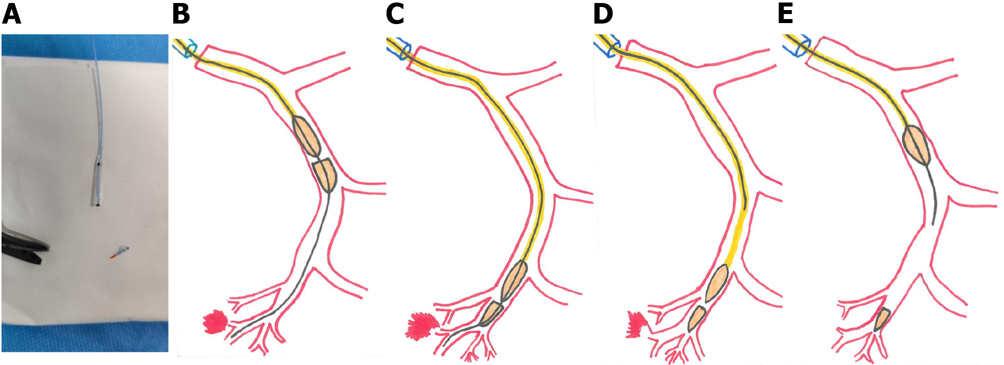
Figure 2 Schematic steps for sealing distal wire perforation using balloon remnant.
A: Distal half of already used balloon is cut by scissors; B and C: The remnant piece is mounted on to the wire in the perforated vessel and pushed with another intact balloon to the distal vessel before the perforation; D and E: Withdrawal of the wire and the intact pushing balloon with the remnant part left distally as the embolization materials.
- Citation: Abdalwahab A, McQuillan C, Farag M, Egred M. Novel economic treatment for coronary wire perforation: A case report. World J Cardiol 2021; 13(6): 177-182
- URL: https://www.wjgnet.com/1949-8462/full/v13/i6/177.htm
- DOI: https://dx.doi.org/10.4330/wjc.v13.i6.177


