Copyright
©The Author(s) 2019.
World J Biol Chem. Jan 7, 2019; 10(1): 17-27
Published online Jan 7, 2019. doi: 10.4331/wjbc.v10.i1.17
Published online Jan 7, 2019. doi: 10.4331/wjbc.v10.i1.17
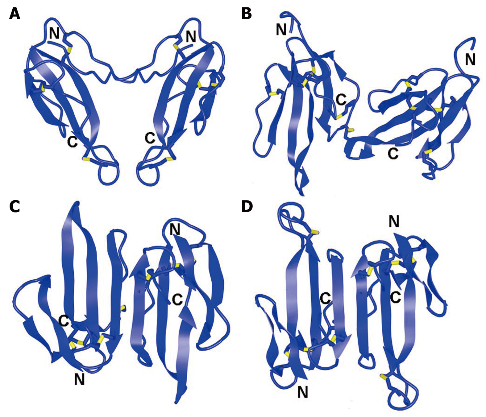
Figure 1 Spatial structures of dimeric three-finger toxins.
A: Homodimer of α-cobratoxin, Protein Data Bank Identification code (PDB ID): 4AEA; B: Irditoxin, PDB ID: 2H7Z; C: Haditoxin, PDB ID: 3HH7; D: κ-bungarotoxin, PDB ID: 1KBA. Disulfide bonds are shown in yellow. N and C indicate N- and C-terminus, respectively.
Figure 2 Alignment of amino acid sequence of toxin Tx7335 with those of nonconventional toxins.
Cysteine residues are marked in yellow. Black lines indicate the locations of the typical disulfide bond in nonconventional toxins; red line indicates the unusual disulfide bond 25-55 in Tx7335. 3NOJ_DENAN: Toxin Tx7335 from Dendroaspis angusticeps (Eastern green mamba); 3NOJ_BUNCA: Bucandin from Bungarus candidus (Malayan krait); 3NOJ6_DENJA: Toxin S6C6 from Dendroaspis jamesoni kaimosae (Eastern Jameson's mamba); 3NOJ_WALAE: Actiflagelin from Walterinnesia aegyptia (desert black snake).
- Citation: Utkin YN. Last decade update for three-finger toxins: Newly emerging structures and biological activities. World J Biol Chem 2019; 10(1): 17-27
- URL: https://www.wjgnet.com/1949-8454/full/v10/i1/17.htm
- DOI: https://dx.doi.org/10.4331/wjbc.v10.i1.17


