Copyright
©2010 Baishideng Publishing Group Co.
World J Biol Chem. Jan 26, 2010; 1(1): 3-12
Published online Jan 26, 2010. doi: 10.4331/wjbc.v1.i1.3
Published online Jan 26, 2010. doi: 10.4331/wjbc.v1.i1.3
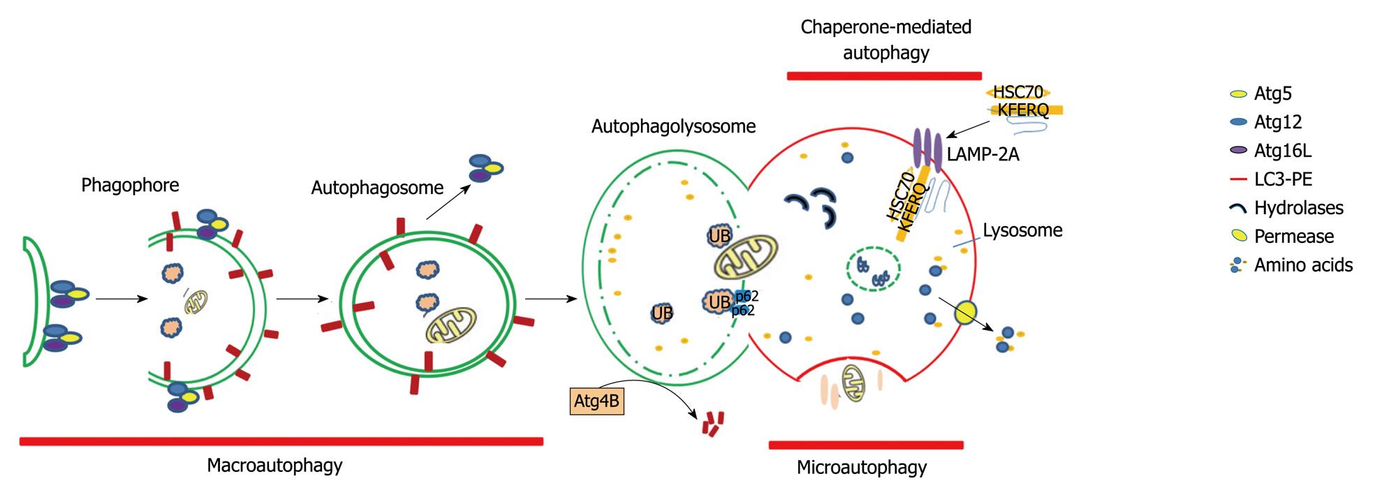
Figure 1 Three forms of autophagy: macroautophagy, microautophagy, and chaperone-mediated autophagy.
Macroautophagy starts with the de novo formation of a cup-shaped isolation membrane or phagophore. The elongation of the isolation membrane is driven by Atg genes while engulfing cytosolic components. The formation of double membrane autophagosomes eventually fuse with lysosomes to form autophagolysosomes where engulfed contents are degraded by lysosomal proteases and hydrolases. Amino acids and other small bio-molecules, such as glucose, are transported back into the cytosol for re-use through the lysosomal membrane permease. Microautophagy involves the engulfment of cytosolic proteins, organelles, and even a piece of nuclear material instantly at the lysosomal membrane by invagination, protrusion, and separation. Chaperone-mediated autophagy is a process of direct transport of a group of proteins that contain a KFERQ motif, which associates with hsc70 and its co-chaperones. This complex then binds with LAMP-2A on the lysosomal membrane. All forms of autophagy subsequently lead to the degradation of intra-autophagosomal components by lysosomal hydrolases. PE: Phosphatidylethanolamine.
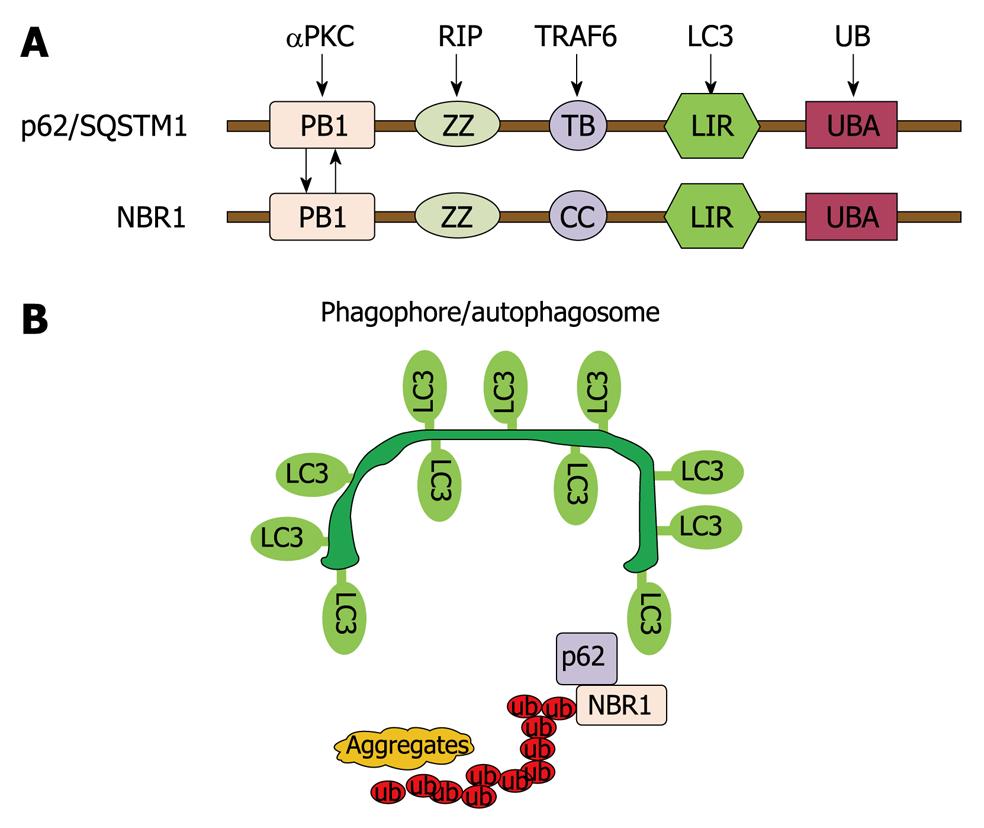
Figure 2 Autophagy regulates protein homeostasis through interaction with p62 and NBR1.
A: A schematic diagram showing the domain organization of p62 and NBR1 proteins. PB1: Phox and Bem1p domain; ZZ: Zinc finger domain; TB: TRAF6-binding domain; CC: Coiled-coil domain; LIR: LC3-interacting region; UBA: Ub-associated domain; B: p62 and NBR1 are autophagy receptors that interact with both ubiquitin-positive protein aggregates through their UBA domains and target them to autophagosomes through their LIR regions with LC3 on the autophagosomal membranes, thereby promoting autophagy of ubiquitinated targets.
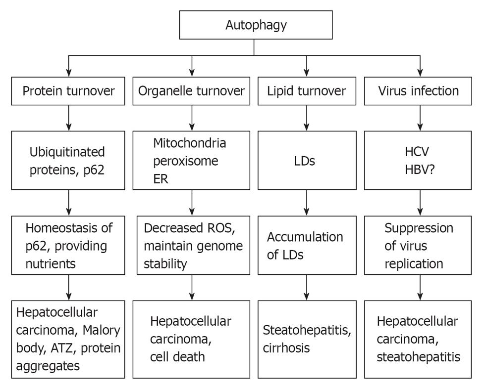
Figure 3 Role of autophagy in liver pathophysiology.
At least 4 different roles that autophagy may play in liver physiology and liver diseases: remove misfolded proteins, regulate hepatocellular organelle turn over, maintain hepatic lipid homeostasis, and influence hepatitis virus infection. As a result, defects in autophagy may lead to accumulation of alcoholic Mallory bodies, α-antitrypsin deficiency-induced liver injury, increased hepatocyte cell death, steatohepatitis and hepatocellular carcinoma. ER: Endoplasmic reticulum; ROS: Reactive oxygen species; LDs: Lipid droplets.
- Citation: Ding WX. Role of autophagy in liver physiology and pathophysiology. World J Biol Chem 2010; 1(1): 3-12
- URL: https://www.wjgnet.com/1949-8454/full/v1/i1/3.htm
- DOI: https://dx.doi.org/10.4331/wjbc.v1.i1.3


