Copyright
©The Author(s) 2016.
World J Gastrointest Surg. Jun 27, 2016; 8(6): 461-466
Published online Jun 27, 2016. doi: 10.4240/wjgs.v8.i6.461
Published online Jun 27, 2016. doi: 10.4240/wjgs.v8.i6.461
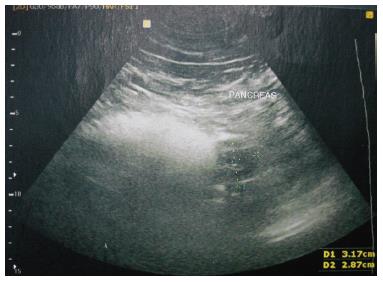
Figure 1 Ultrasound echo showing a 2.
8 cm × 3.1 cm hypoechoic solid mass located on the pancreatic body.
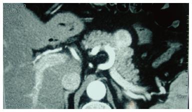
Figure 2 Computed tomography scan revealing a 3.
9 cm exophytic mass, arising from the pancreatic body, which was enhanced from the arterial to portal venous phase.
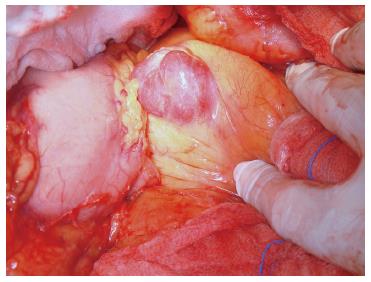
Figure 3 Intra-operative appearance of the pancreatic mass.
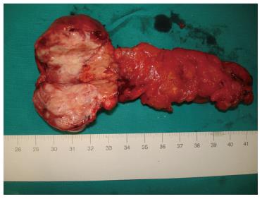
Figure 4 Pancreatic mass specimen.
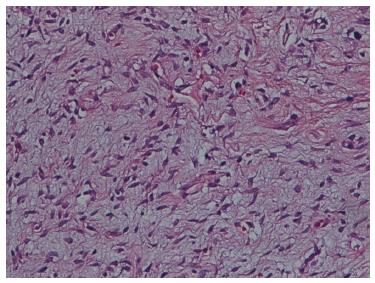
Figure 5 Solitary fibrous tumor with a hyalinized area, HE stain.
Magnification: × 400 .
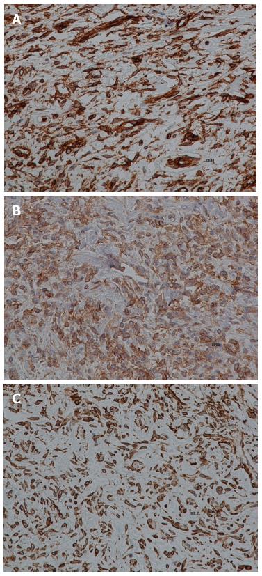
Figure 6 Immunohistochemical staining (brown) for CD34 (A), CD99 (B) and Bcl-2 (C).
Magnification: × 400.
- Citation: Paramythiotis D, Kofina K, Bangeas P, Tsiompanou F, Karayannopoulou G, Basdanis G. Solitary fibrous tumor of the pancreas: Case report and review of the literature. World J Gastrointest Surg 2016; 8(6): 461-466
- URL: https://www.wjgnet.com/1948-9366/full/v8/i6/461.htm
- DOI: https://dx.doi.org/10.4240/wjgs.v8.i6.461


