Copyright
©The Author(s) 2015.
World J Gastrointest Surg. Apr 27, 2015; 7(4): 67-70
Published online Apr 27, 2015. doi: 10.4240/wjgs.v7.i4.67
Published online Apr 27, 2015. doi: 10.4240/wjgs.v7.i4.67
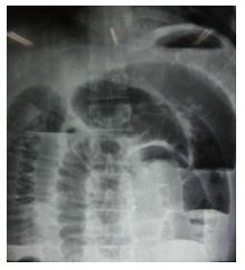
Figure 1 Abdominal radiograph showing multiple distended loops of small bowel with fluid levels.
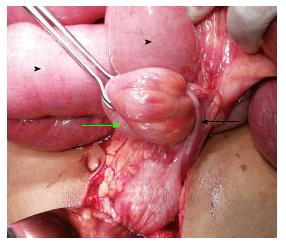
Figure 2 The Appendix (black arrow) encircling the loop of terminal Ileum (green arrow) with dilatation of proximal small bowel (black arrowheads).
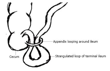
Figure 3 Depiction of Appendix wrapping around loop of Ileum.
(Reproduced with permision from Menon et al[7]).
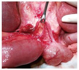
Figure 4 Inflamed and odematous tip of Appendix adherent to the root of mesentry (black arrow).
- Citation: Awale L, Joshi BR, Rajbanshi S, Adhikary S. Appendiceal tie syndrome: A very rare complication of a common disease. World J Gastrointest Surg 2015; 7(4): 67-70
- URL: https://www.wjgnet.com/1948-9366/full/v7/i4/67.htm
- DOI: https://dx.doi.org/10.4240/wjgs.v7.i4.67


