Copyright
©The Author(s) 2015.
World J Gastrointest Surg. Oct 27, 2015; 7(10): 273-278
Published online Oct 27, 2015. doi: 10.4240/wjgs.v7.i10.273
Published online Oct 27, 2015. doi: 10.4240/wjgs.v7.i10.273
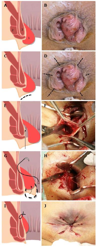
Figure 1 Sagittal diagrams (A, C, E, G and I) and intraoperative views (B, D, F, H and J) of the anus.
Small incisions were made in the perianal skin (C), and several radial incisions were performed (D). The tissue between the internal sphincter muscle and anal cushion was dissected (E and F). The anal cushion was restored to its native position (G). The anal cushion was sutured from its middle portion to the cranial side using single stitches (G and H). All sutures were tied up circumferentially (I). The appearance of the anus after the completion of the ACL procedure (J). 1: Hemorrhoids; 2: External sphincter; 3: Internal sphincter. Dotted line: Incision; Arrow: Dissection.
- Citation: Ishiyama G, Nishidate T, Ishiyama Y, Nishio A, Tarumi K, Kawamura M, Okita K, Mizuguchi T, Fujimiya M, Hirata K. Anal cushion lifting method is a novel radical management strategy for hemorrhoids that does not involve excision or cause postoperative anal complications. World J Gastrointest Surg 2015; 7(10): 273-278
- URL: https://www.wjgnet.com/1948-9366/full/v7/i10/273.htm
- DOI: https://dx.doi.org/10.4240/wjgs.v7.i10.273


