Copyright
©2014 Baishideng Publishing Group Inc.
World J Gastrointest Surg. Aug 27, 2014; 6(8): 156-159
Published online Aug 27, 2014. doi: 10.4240/wjgs.v6.i8.156
Published online Aug 27, 2014. doi: 10.4240/wjgs.v6.i8.156
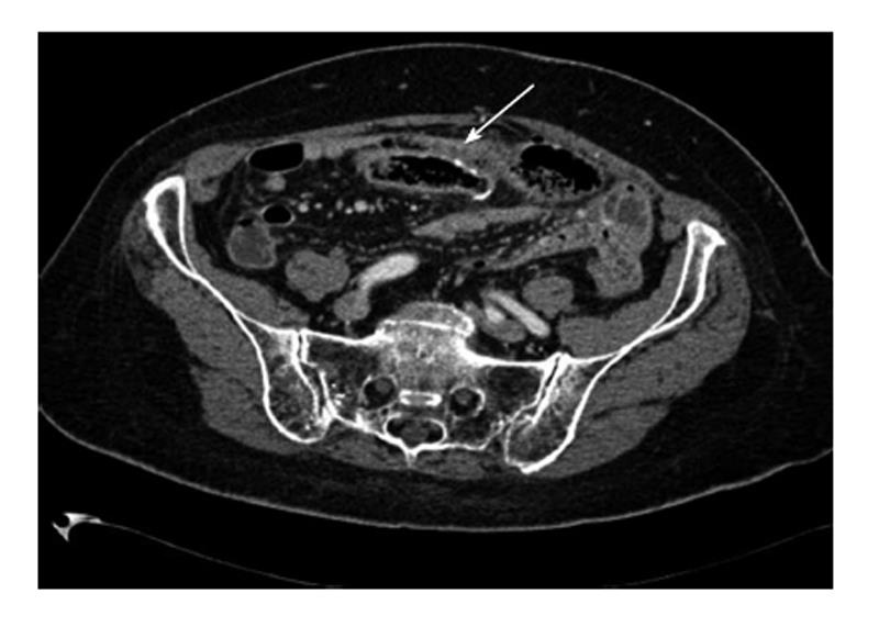
Figure 1 Computed tomography.
The scan shows intraperitoneal free air with air bubbles around the ileocolonic anastomosis and a dilated blind loop with thickened bowel walls and mucosal hyperemia.
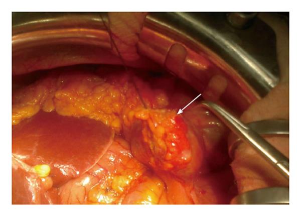
Figure 2 The image shows a long blind loop with signs of microperforation on the suture line.
- Citation: Dalla Valle R, Zinicola R, Iaria M. Blind loop perforation after side-to-side ileocolonic anastomosis. World J Gastrointest Surg 2014; 6(8): 156-159
- URL: https://www.wjgnet.com/1948-9366/full/v6/i8/156.htm
- DOI: https://dx.doi.org/10.4240/wjgs.v6.i8.156


