Copyright
©2014 Baishideng Publishing Group Inc.
World J Gastrointest Surg. Jun 27, 2014; 6(6): 107-111
Published online Jun 27, 2014. doi: 10.4240/wjgs.v6.i6.107
Published online Jun 27, 2014. doi: 10.4240/wjgs.v6.i6.107
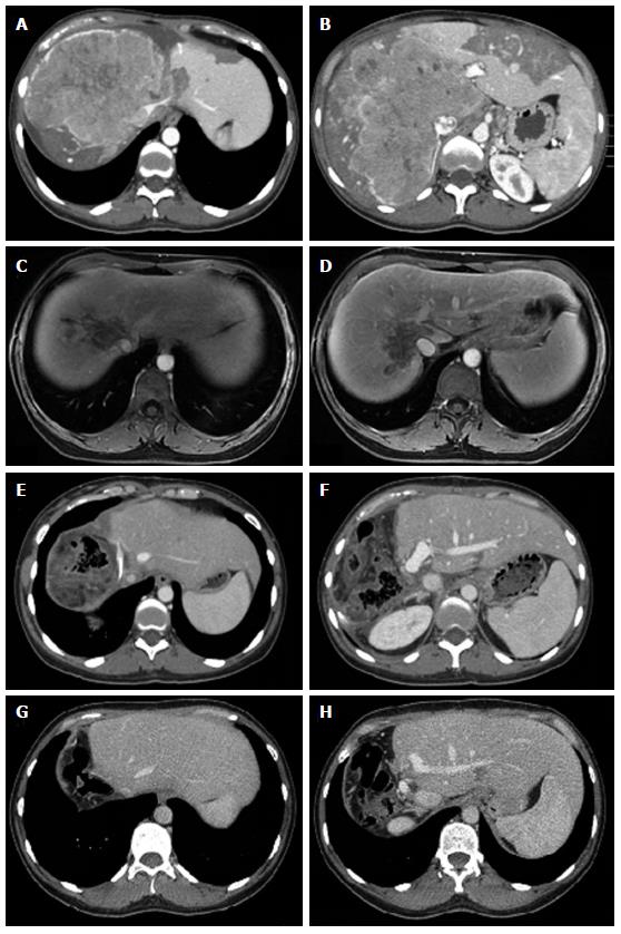
Figure 1 Abdominal computed tomography and magnetic resonance imaging of a 35-year-old Caucasian female, with no previous history of liver disease, showing a large mass in the upper right quadrant.
A, B: Abdominal computed tomography (CT) before chemotherapy showing a large mass invading (17 cm × 15 cm) the right and the middle hepatic veins, and surrounding the left hepatic vein; C, D: Abdominal magnetic resonance imaging after gemcitabine-oxaliplatin chemotherapy showing significant reduction of the tumor size; E, F: Abdominal CT two weeks after surgery with hypertrophy of the left lateral segment of liver (liver remnant), and free and patent left branch of the portal vein and left hepatic vein; G, H: Late follow-up after 14 mo.
- Citation: Fonseca GM, Varella AD, Coelho FF, Abe ES, Dumarco RB, Herman P. Downstaging and resection after neoadjuvant therapy for fibrolamellar hepatocellular carcinoma. World J Gastrointest Surg 2014; 6(6): 107-111
- URL: https://www.wjgnet.com/1948-9366/full/v6/i6/107.htm
- DOI: https://dx.doi.org/10.4240/wjgs.v6.i6.107


