Copyright
©2014 Baishideng Publishing Group Inc.
World J Gastrointest Surg. May 27, 2014; 6(5): 80-83
Published online May 27, 2014. doi: 10.4240/wjgs.v6.i5.80
Published online May 27, 2014. doi: 10.4240/wjgs.v6.i5.80
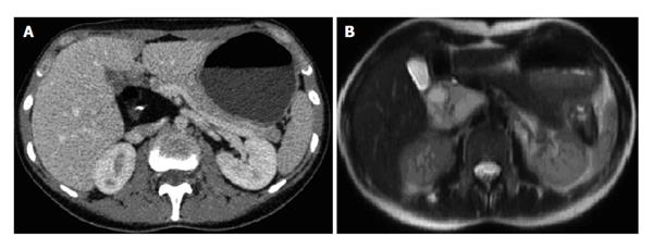
Figure 1 Cross-sectional imaging.
A hepatoduodenal heterogeneous mass was revealed by A: Computed tomography; B: Magnetic resonance imaging.
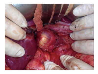
Figure 2 Operative finding.
A laparotomy revealed an encapsulated lesion without invasion to adjacent organs or vessels (1: Common bile duct; 2: Teratoma; 3: Right lobe of the liver).
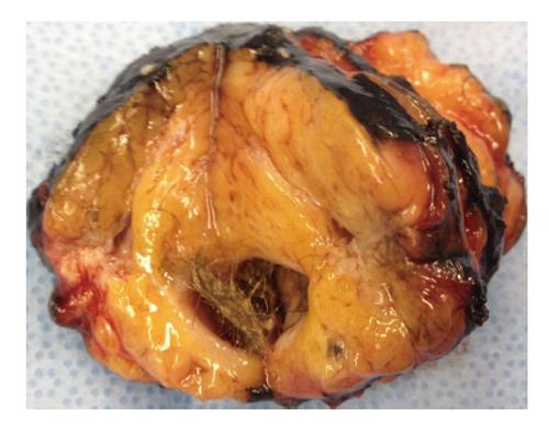
Figure 3 Tumor appearance.
The resected heterogeneous lesion was composed of fat tissue, calcifications and hair.
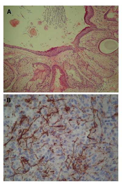
Figure 4 Histopathology of the tumor.
Microscopic examination of the specimen revealed A: A cystic wall with cutaneous annexes; B: Glial fibrillary acidic protein immunoreactivity.
- Citation: Jeismann VB, Dumarco RB, Loreto CD, Barbuti RC, Jukemura J. Rare cause of abdominal incidentaloma: Hepatoduodenal ligament teratoma. World J Gastrointest Surg 2014; 6(5): 80-83
- URL: https://www.wjgnet.com/1948-9366/full/v6/i5/80.htm
- DOI: https://dx.doi.org/10.4240/wjgs.v6.i5.80


