Copyright
©2013 Baishideng Publishing Group Co.
World J Gastrointest Surg. Jun 27, 2013; 5(6): 173-177
Published online Jun 27, 2013. doi: 10.4240/wjgs.v5.i6.173
Published online Jun 27, 2013. doi: 10.4240/wjgs.v5.i6.173
Figure 1 Differences in the view and approach between open and laparoscopic liver surgery.
An illustration depicting the differences between the different surgery types. Since the view of operative field is in caudal-to-cranial direction in the laparoscopic procedure (LAP, right), not anterior-to-posterior (dorsal) as in the open procedure (OPEN, left), there is an advantage of good vision for the dorsal part of the liver and inferior vena cava. IVC: Inferior vena cava.
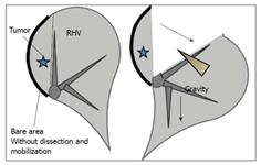
Figure 2 A schematic diagram of the caudal approach of pure laparoscopic posterior sectionectomy without mobilization under laparoscopy-specific view.
Since the remnant liver sinks down and the resected liver is fixed to retroperitoneum, the cutting surface is well-opened and the exposure of right hepatic vein (RHV) is facilitated (arrow). Transection of the liver and exposure of the right hepatic vein, dividing its posterior branches one by one, proceed in one direction from caudal to cranial, simultaneously with the dissection of inferior vena cava anterior wall.
Figure 3 Computed tomography scan findings.
A 63-year-old woman with a 1.5 cm metachronous colorectal liver metastasis in the segment 6 close to the right hepatic vein underwent adjuvant chemotherapy after open colorectal surgery and her liver had severe fatty change. Therefore, posterior sectionectomy was applied to her lesion, not right hepatectomy. However, since the lesion is close to the right hepatic vein (RHV), RHV exposure was required for R0 resection.
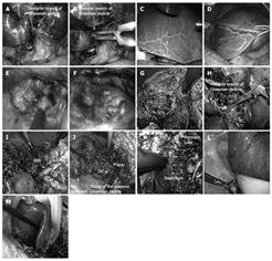
Figure 4 Intraoperative findings of the case.
A-D: Part I; E-H: Part II; I, J: Part III; K-M: Part IV. After cholecystectomy, Glissonian pedicle of the right lobe and posterior sector was encircled (A). The posterior pedicle was clamped (B), allowing the resection line to be visualized as; an ischemic demarcation line (C, D). E, F: The anterior wall of the inferior vena cava was dissected behind the liver; G: Liver parenchyma was transected on the demarcation line, and the peripheral part of the right hepatic vein was exposed, clipped, and divided; H: After the cutting line reached the level of Glissonian, the posterior pedicle was divided with a linear stapler. Transection of the liver and the exposure of the right hepatic vein (RHV) by dividing its posterior branched one-by-one proceeded in one direction from caudal to cranial, simultaneously with the dissection of the inferior vena cava anterior wall. Since remnants of the liver sunk down and the resected liver was fixed to the retroperitoneum, the cutting surface was well opened and the exposure of the RHV was facilitated. K: Following completion of the liver parenchymal transection; L: Dissection of the resected liver from retroperitoneum was performed; M: The resected liver was removed. Dissection of the resected liver from retroperitoneum was easily performed with the use of gravity. The operation time was 341 min and operative blood loss was 1356 mL. The patient’s postoperative hospital stay was uneventful and she was well without any signs of recurrences one year and two months after surgery.
- Citation: Tomishige H, Morise Z, Kawabe N, Nagata H, Ohshima H, Kawase J, Arakawa S, Yoshida R, Isetani M. Caudal approach to pure laparoscopic posterior sectionectomy under the laparoscopy-specific view. World J Gastrointest Surg 2013; 5(6): 173-177
- URL: https://www.wjgnet.com/1948-9366/full/v5/i6/173.htm
- DOI: https://dx.doi.org/10.4240/wjgs.v5.i6.173


