Copyright
©2013 Baishideng Publishing Group Co.
World J Gastrointest Surg. Feb 27, 2013; 5(2): 16-21
Published online Feb 27, 2013. doi: 10.4240/wjgs.v5.i2.16
Published online Feb 27, 2013. doi: 10.4240/wjgs.v5.i2.16
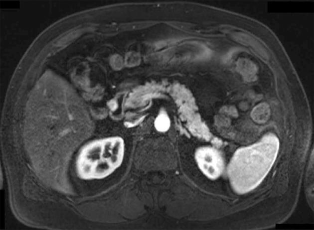
Figure 1 magnetic resonance scan of small gastrointestinal stromal tumors in the second portion of the duodenum resected by laparoscopic wedge resection (Patient 3).
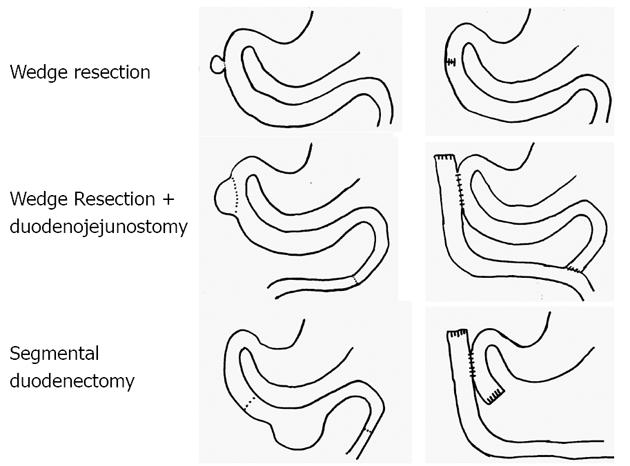
Figure 2 Illustration of the different techniques for limited resection of duodenal gastrointestinal stromal tumors.
Wedge resection was performed for small tumors and for larger tumors with only a small-area base at the duodenal wall in all parts of the duodenum. Wedge resection with reconstruction of the defect at the duodenal wall by duodenojejunostomy was performed in patients with larger defects of the second part of the duodenum after tumor resection. Segmental duodenectomy was carried out for larger tumors of the third or fourth part of the duodenum.
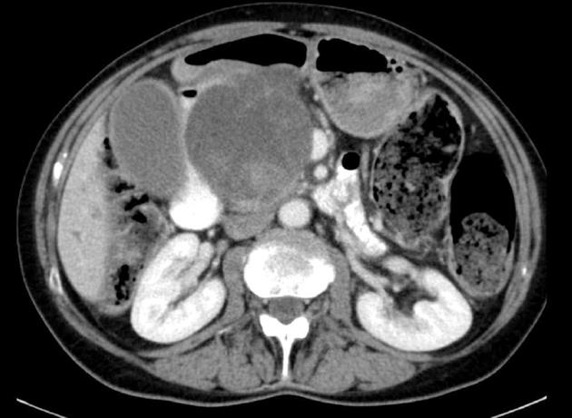
Figure 3 Computed tomography scan of a high-risk large duodenal gastrointestinal stromal tumors which hast been resected by pancreatoduodenectomy including portal vein resection (Patient 4).
The origin of the tumor is located in the first part of the duodenum were pathology revealed a 65 mm x 55 mm ulcer.
- Citation: Hoeppner J, Kulemann B, Marjanovic G, Bronsert P, Hopt UT. Limited resection for duodenal gastrointestinal stromal tumors: Surgical management and clinical outcome. World J Gastrointest Surg 2013; 5(2): 16-21
- URL: https://www.wjgnet.com/1948-9366/full/v5/i2/16.htm
- DOI: https://dx.doi.org/10.4240/wjgs.v5.i2.16


