Copyright
©2013 Baishideng Publishing Group Co.
World Journal of Gastrointestinal Surgery. Nov 27, 2013; 5(11): 309-313
Published online Nov 27, 2013. doi: 10.4240/wjgs.v5.i11.309
Published online Nov 27, 2013. doi: 10.4240/wjgs.v5.i11.309
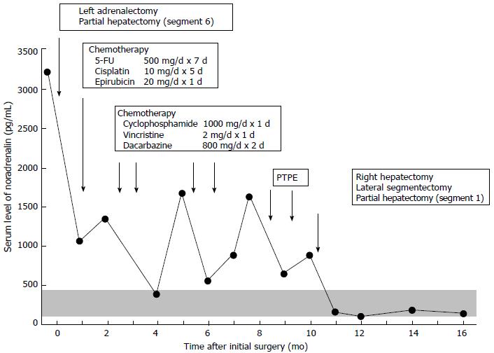
Figure 1 Serum catecholamine levels before and after treatments.
Serum catecholamine levels are shown in relation to surgery, chemotherapy and preoperative percutaneous transhepatic portal vein embolization. The shaded area represents the normal range (100-450 pg/mL). PTPE: Percutaneous transhepatic portal.
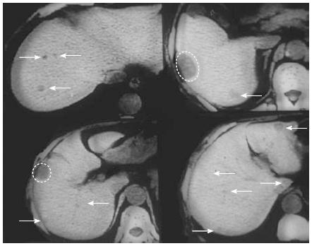
Figure 2 Angio-computed tomography findings.
Preoperative image study revealed multiple liver metastases (white arrows) except for segment 4. She underwent the partial hepatectomy of segment 6 at initial surgery (white dotted circle).
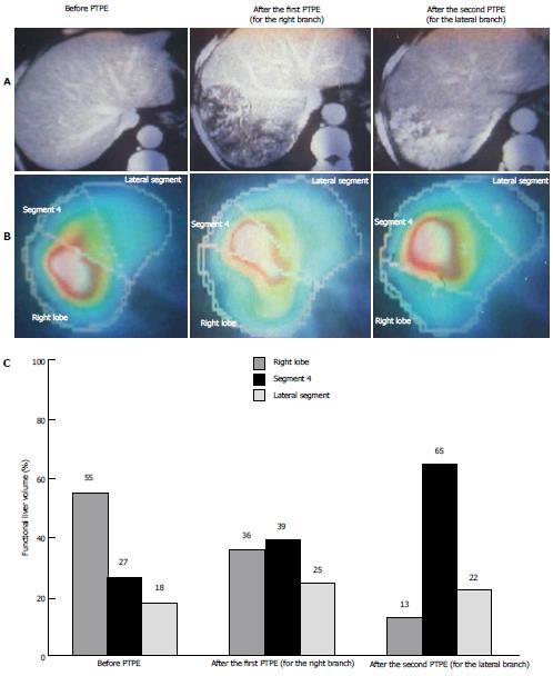
Figure 3 Changes in functional hepatic volume after percutaneous transhepatic portal vein embolization.
The angio-computed tomography findings (A), technetium-99m diethylenetriamine pentaacetic acid galactosyl human serum albumin scintigraphy for asialoglycoprotein receptor (B) and the functional hepatic volume (C) were shown in each lobe/segment before and after percutaneous transhepatic portal vein embolization. Functional hepatic volume of each lobe/segment was calculated as a percentage of whole liver volume. PTPE: Percutaneous transhepatic portal.
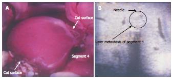
Figure 4 Intraoperative findings.
A: She underwent right hepatectomy with lateral segmentectomy and partial resection of segment 1. B: Intraoperative ultrasonography detected two small nodules in segment 4, which were treated with intraoperative microwave coagulation therapy.
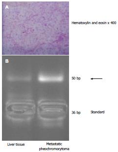
Figure 5 Histopathological finding and telomerase activity in the resected tumor.
A: Histopathological examination of the surgical specimens was consistent with pheochromocytoma; B: Real-time polymerase chain reaction clearly revealed increased telomerase activity (arrow) in the resected tumor tissue compared with liver tissue.
- Citation: Hori T, Yamagiwa K, Hayashi T, Yagi S, Iida T, Taniguchi K, Kawarada Y, Uemoto S. Malignant pheochromocytoma: Hepatectomy for liver metastases. World Journal of Gastrointestinal Surgery 2013; 5(11): 309-313
- URL: https://www.wjgnet.com/1948-9366/full/v5/i11/309.htm
- DOI: https://dx.doi.org/10.4240/wjgs.v5.i11.309


