Copyright
©2012 Baishideng Publishing Group Co.
World J Gastrointest Surg. Aug 27, 2012; 4(8): 203-207
Published online Aug 27, 2012. doi: 10.4240/wjgs.v4.i8.203
Published online Aug 27, 2012. doi: 10.4240/wjgs.v4.i8.203
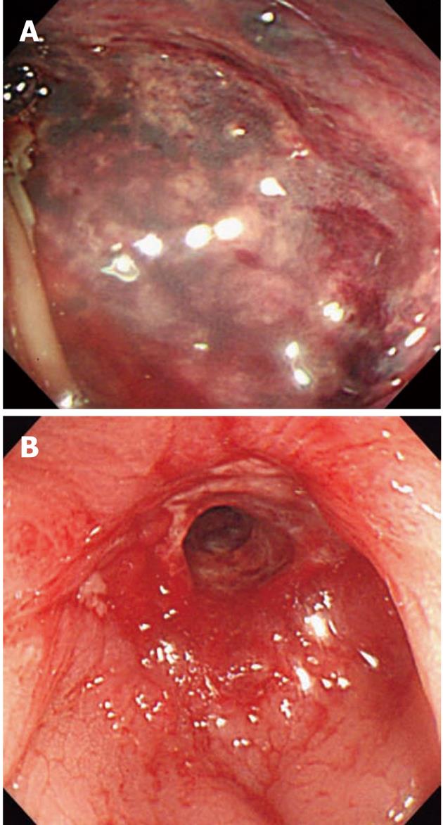
Figure 1 Endoscopic findings.
A: Initial endoscopic findings, with entire mucosal deficiency and oozing at 20 cm from the anal verge; B: Strictured colon wall after conservative treatment.
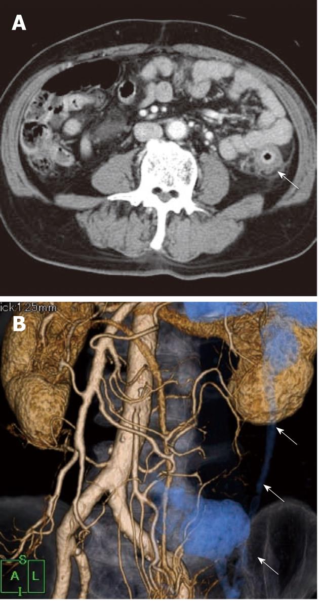
Figure 2 Computed tomography and 3D reconstituted imaging.
A: Computed tomography shows the strictured descending colon wall (arrow); B: Strictured colon for a length of 15 cm (arrows).
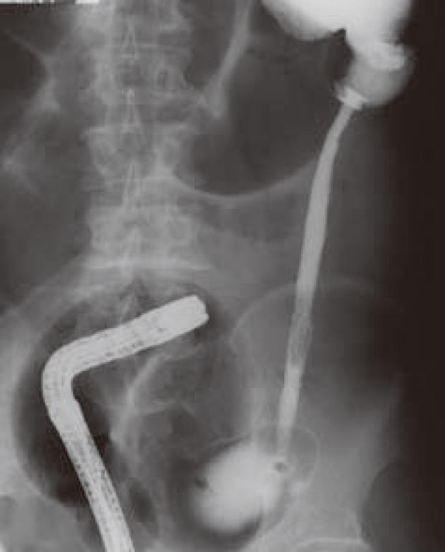
Figure 3 Radiographic contrast study of the lower gastrointestinal tract.
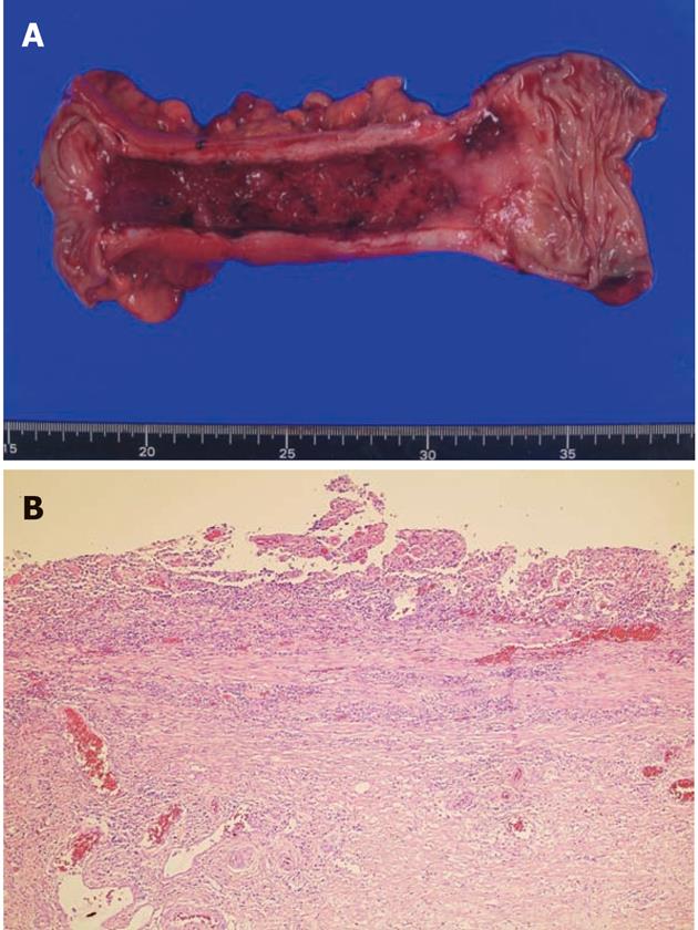
Figure 4 Laparoscopy-assisted left colectomy.
A: Macroscopic appearance of the resected specimen; B: Histopathological findings of the strictured colon (HE, staining × 40).
- Citation: Tsukada T, Nakano T, Matsui D, Sasaki S. Stenotic ischemic colitis treated with laparoscopy-assisted surgery. World J Gastrointest Surg 2012; 4(8): 203-207
- URL: https://www.wjgnet.com/1948-9366/full/v4/i8/203.htm
- DOI: https://dx.doi.org/10.4240/wjgs.v4.i8.203


