Copyright
©2012 Baishideng Publishing Group Co.
World J Gastrointest Surg. Aug 27, 2012; 4(8): 199-202
Published online Aug 27, 2012. doi: 10.4240/wjgs.v4.i8.199
Published online Aug 27, 2012. doi: 10.4240/wjgs.v4.i8.199
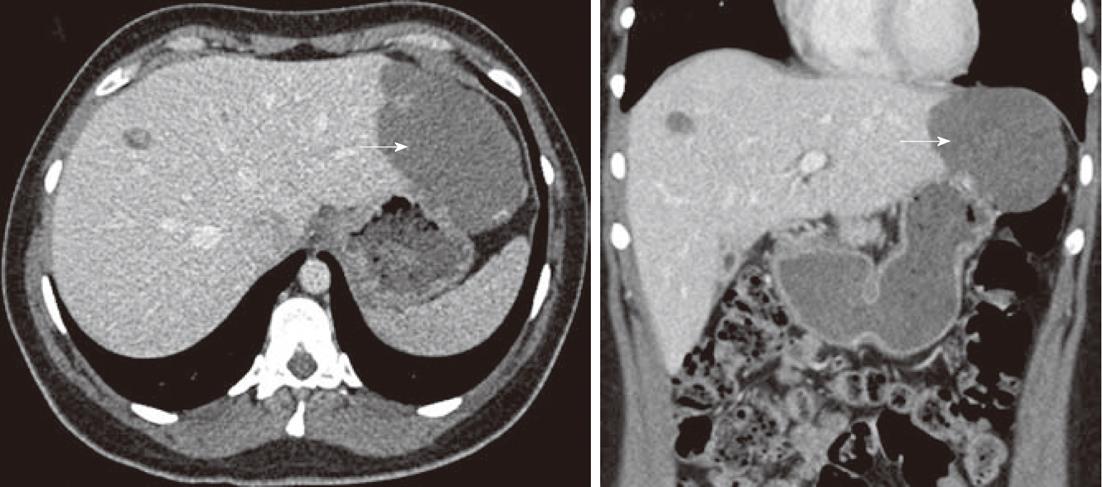
Figure 1 Axial section (A) and coronal section (B) of enhanced portal-phase computed tomography scan showing multiple haemangiomas of the left and right liver lobes, the largest (arrow) in segments 2 and 3 measuring 10 cm in diameter.
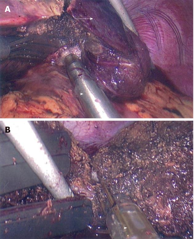
Figure 2 Laparoscopic resection of a giant haemangioma (A, arrow) using the laparoscopic Habib™ 4× bipolar resection device (B, arrowhead).
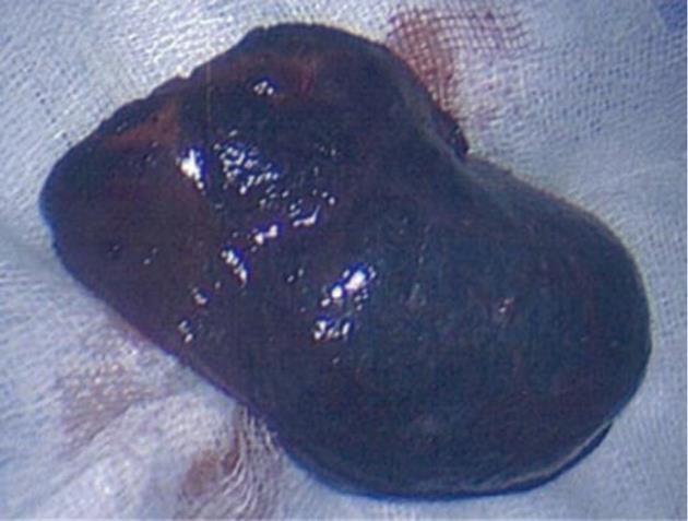
Figure 3 Ten cm giant haemangioma following laparoscopic resection.
- Citation: Acharya M, Panagiotopoulos N, Bhaskaran P, Kyriakides C, Pai M, Habib N. Laparoscopic resection of a giant exophytic liver haemangioma with the laparoscopic Habib 4× radiofrequency device. World J Gastrointest Surg 2012; 4(8): 199-202
- URL: https://www.wjgnet.com/1948-9366/full/v4/i8/199.htm
- DOI: https://dx.doi.org/10.4240/wjgs.v4.i8.199


