Copyright
©2011 Baishideng Publishing Group Co.
World J Gastrointest Surg. Apr 27, 2011; 3(4): 54-55
Published online Apr 27, 2011. doi: 10.4240/wjgs.v3.i4.54
Published online Apr 27, 2011. doi: 10.4240/wjgs.v3.i4.54
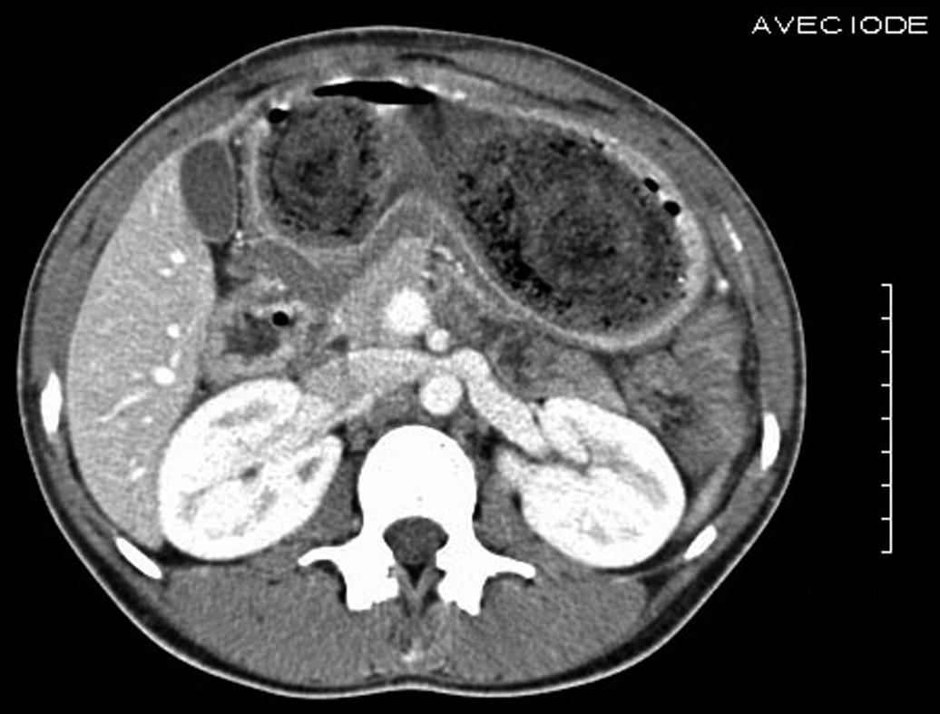
Figure 1 The trichobezoar presented as a heterogenous non-enhancing mass in the stomach on computed tomography scan with IV contrast.
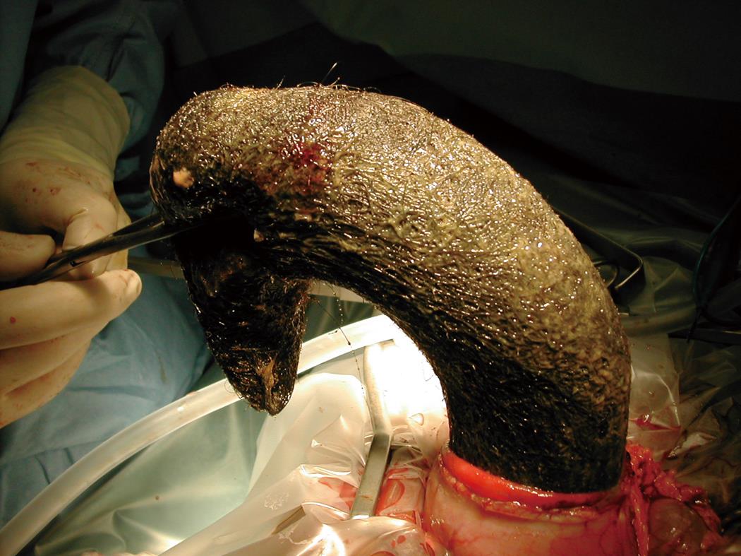
Figure 2 Intraoperative view of the trichobezoar and its extraction through a gastrotomy.
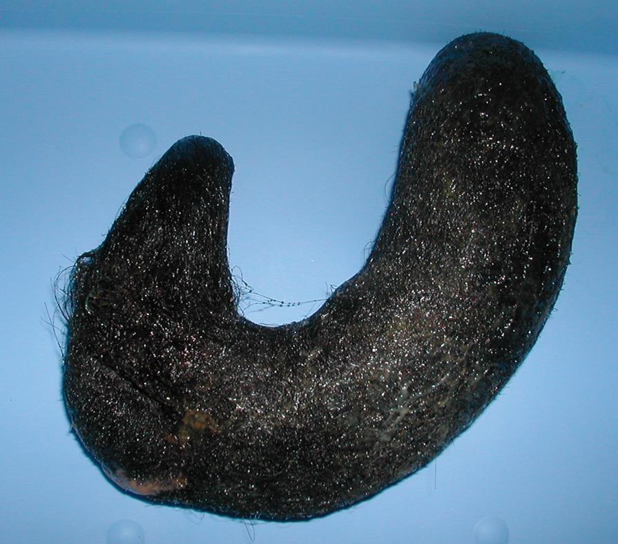
Figure 3 Gross picture of the trichobezoar.
- Citation: Gaujoux S, Bach G, Au J, Godiris-Petit G, Munoz-Bongrand N, Cattan P, Sarfati E. Trichobezoar: A rare cause of bowel obstruction. World J Gastrointest Surg 2011; 3(4): 54-55
- URL: https://www.wjgnet.com/1948-9366/full/v3/i4/54.htm
- DOI: https://dx.doi.org/10.4240/wjgs.v3.i4.54


