Copyright
©2010 Baishideng.
World J Gastrointest Surg. Jun 27, 2010; 2(6): 187-192
Published online Jun 27, 2010. doi: 10.4240/wjgs.v2.i6.187
Published online Jun 27, 2010. doi: 10.4240/wjgs.v2.i6.187
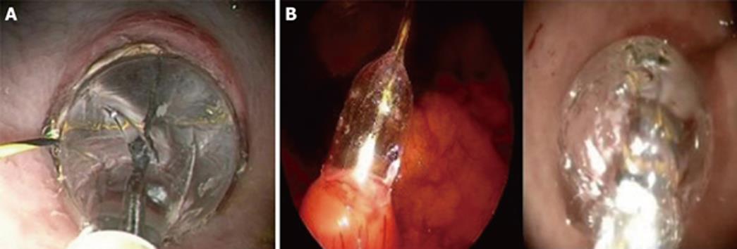
Figure 1 Balloon dilatation of the gastrotomy in the animal model (A) and in a patient (B).
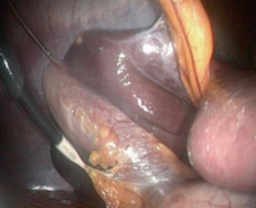
Figure 2 Retraction of the gallbladder with a laparoscopic micro-instrument introduced in the right hypochondrium.
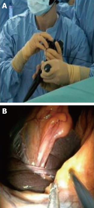
Figure 3 Dissection of the cystic pedicle with a flexible endoscope: “four hands” technique.
Extrernal view (A) and endoscopic view (B).
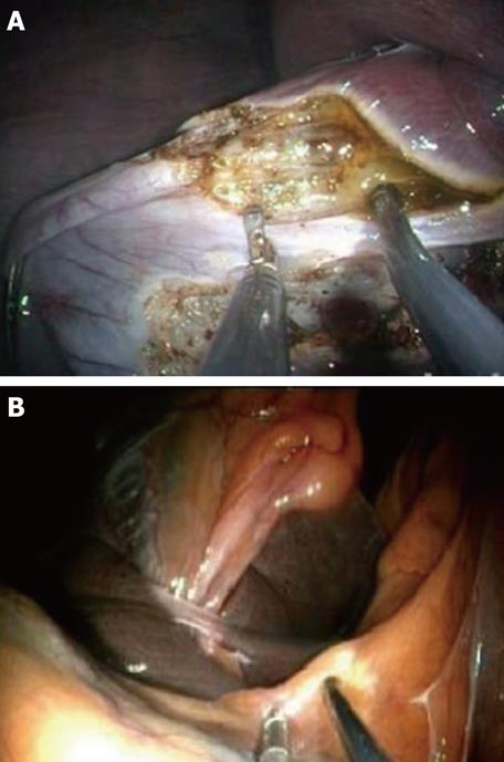
Figure 4 Dissection of the cystic pedicle with a flexible endoscope in the animal model (A) and in a patient (B).
- Citation: Dallemagne B, Perretta S, Allemann P, Donatelli G, Asakuma M, Mutter D, Marescaux J. Transgastric cholecystectomy: From the laboratory to clinical implementation. World J Gastrointest Surg 2010; 2(6): 187-192
- URL: https://www.wjgnet.com/1948-9366/full/v2/i6/187.htm
- DOI: https://dx.doi.org/10.4240/wjgs.v2.i6.187


