Copyright
©2010 Baishideng.
World J Gastrointest Surg. Mar 27, 2010; 2(3): 89-94
Published online Mar 27, 2010. doi: 10.4240/wjgs.v2.i3.89
Published online Mar 27, 2010. doi: 10.4240/wjgs.v2.i3.89
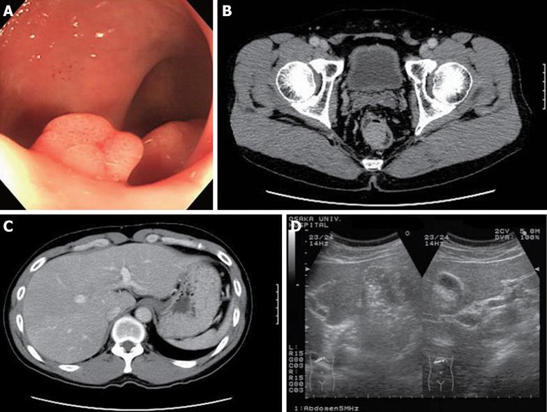
Figure 1 Preoperative colonoscopy and image examinations.
A: Colonoscopy showed an elevated yellowish lesion with a slight central depression of which size was 30 mm in diameter, in the lower rectum; B: A tumor was present on the right wall of the lower rectum by computed tomography (CT) scan; C: Abdominal CT failed to show any obvious abnormalities in the liver; D: No obvious lesions were detected in the liver by ultrasonography.
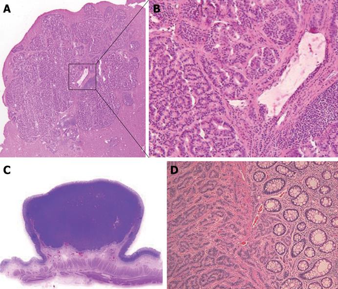
Figure 2 Histopathology of liver metastasis and primary tumor.
A: Histopathology of a hepatic nodule (Magnification: × 20). A section was stained by hematoxiline & eosin (H&E) solution; B: Magnified image of (A) (Magnification: × 100): The tumor cells were homogeneous and spherical, forming ribbon-like structures, which was compatible with the carcinoid tumor; C: Resected rectal tumor. A loupe image; D: Histopathology of the rectal tumor (Magnification: × 100): Identical histopathological features to that of liver metastasis were confirmed.
- Citation: Yamamoto H, Hemmi H, Gu JY, Sekimoto M, Doki Y, Mori M. Minute liver metastases from a rectal carcinoid: A case report and review. World J Gastrointest Surg 2010; 2(3): 89-94
- URL: https://www.wjgnet.com/1948-9366/full/v2/i3/89.htm
- DOI: https://dx.doi.org/10.4240/wjgs.v2.i3.89


