Copyright
©2010 Baishideng Publishing Group Co.
World J Gastrointest Surg. Nov 27, 2010; 2(11): 385-388
Published online Nov 27, 2010. doi: 10.4240/wjgs.v2.i11.385
Published online Nov 27, 2010. doi: 10.4240/wjgs.v2.i11.385
Figure 1 Endoscopy, endoscopic ultrasound and computed tomography findings of the patient.
A: Endoscopy revealed a submucosal tumor (arrow) without central depression on the posterior of the antrum; B: Endoscopic ultrasound revealed a tumor (arrow) arising from the muscle layer; C: Computed tomography scan showed the tumor (arrow) stained in the early phase.
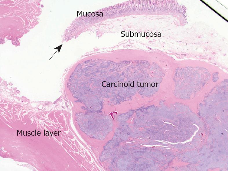
Figure 2 Resected specimen viewed using low magnification.
The carcinoid tumors were located in the submucosal layer infiltrating the muscle layer. The muscularis mucosa (arrow) was intact.
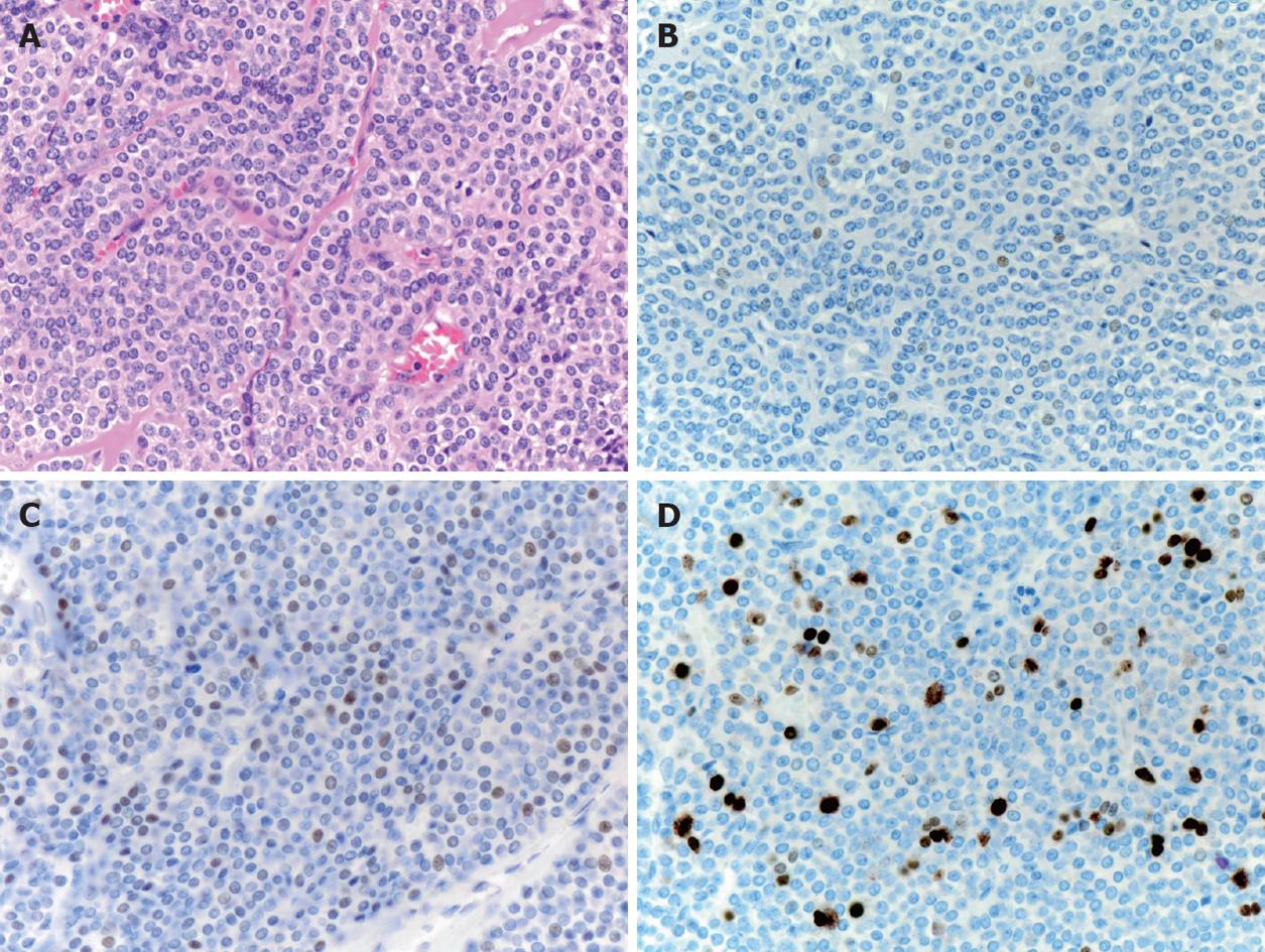
Figure 3 Pathological findings of the resected carcinoid (× 40).
A: Hematoxylin and eosin; B: Chromogranin A staining; C: P53 staining; D: Ki-67 staining.
- Citation: Kinoshita T, Oshiro T, Urita T, Yoshida Y, Ooshiro M, Okazumi S, Katoh R, Sasai D, Hiruta N. Sporadic gastric carcinoid tumor successfully treated by two-stage laparoscopic surgery: A case report. World J Gastrointest Surg 2010; 2(11): 385-388
- URL: https://www.wjgnet.com/1948-9366/full/v2/i11/385.htm
- DOI: https://dx.doi.org/10.4240/wjgs.v2.i11.385


