
















Copyright: ©Author(s) 2026.
World J Gastrointest Surg. Nov 27, 2026; 18(11): 125509
Published online Nov 27, 2026. doi: 10.4240/wjgs.125509
Published online Nov 27, 2026. doi: 10.4240/wjgs.125509
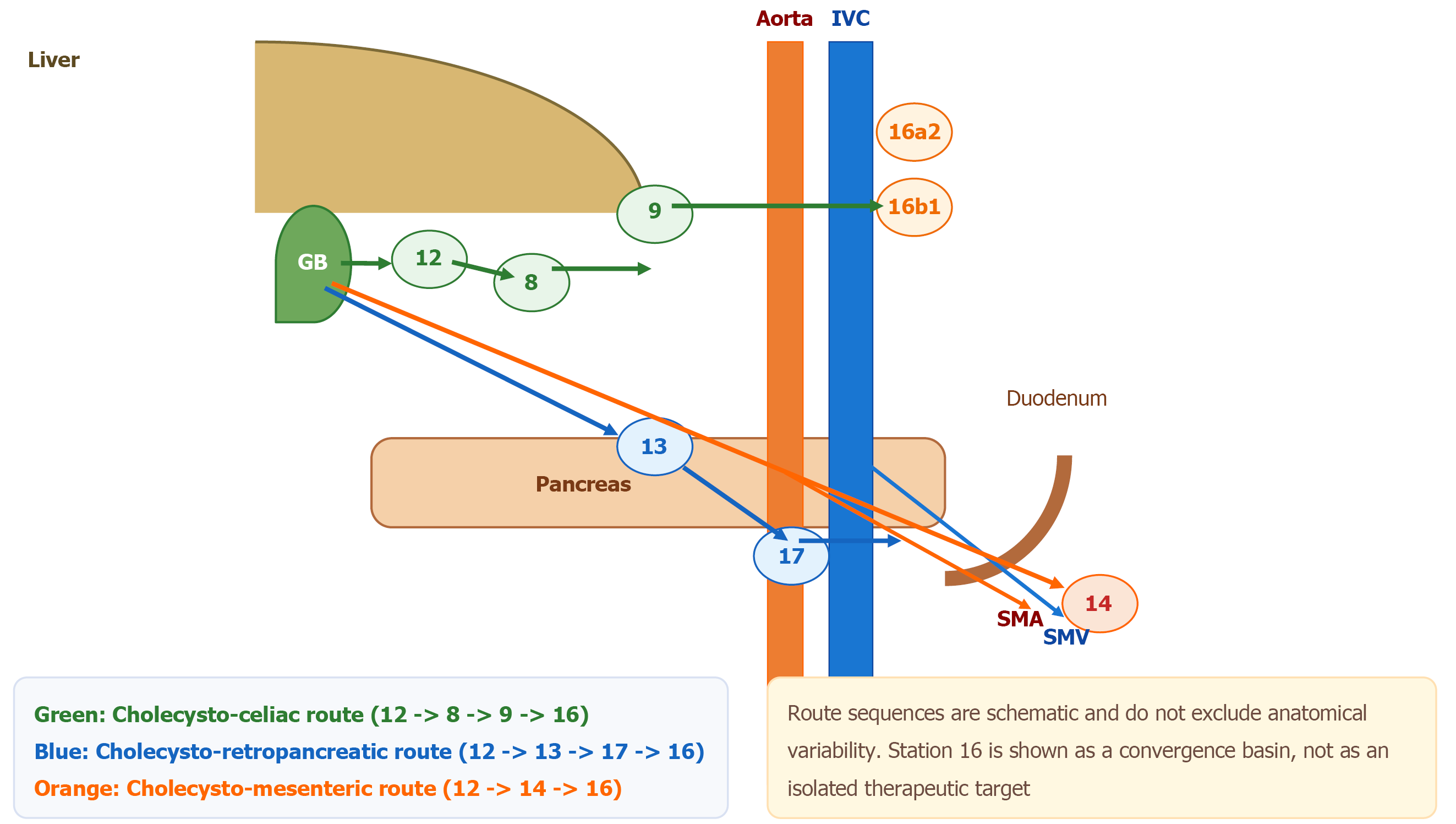
Figure 1 Semi-anatomical map of the three lymphatic pathways of gallbladder cancer.
The cholecysto-celiac pathway (green) involves stations 12, 8 and 9; the cholecysto-retropancreatic pathway (blue) involves stations 12, 13 and 17; the cholecysto-mesenteric pathway (orange) involves station 14 along the superior mesenteric vessels. All pathways converge toward station 16, particularly 16a2 and 16b1. This is an original schematic prepared by the authors based on classical lymphatic studies and Japanese Society of Biliary Surgery station nomenclature[4-6]. The route sequences are simplified and should not be interpreted as universally fixed anatomy. IVC: Inferior vena cava; SMA: Superior mesenteric artery; SMV: Superior mesenteric vein.
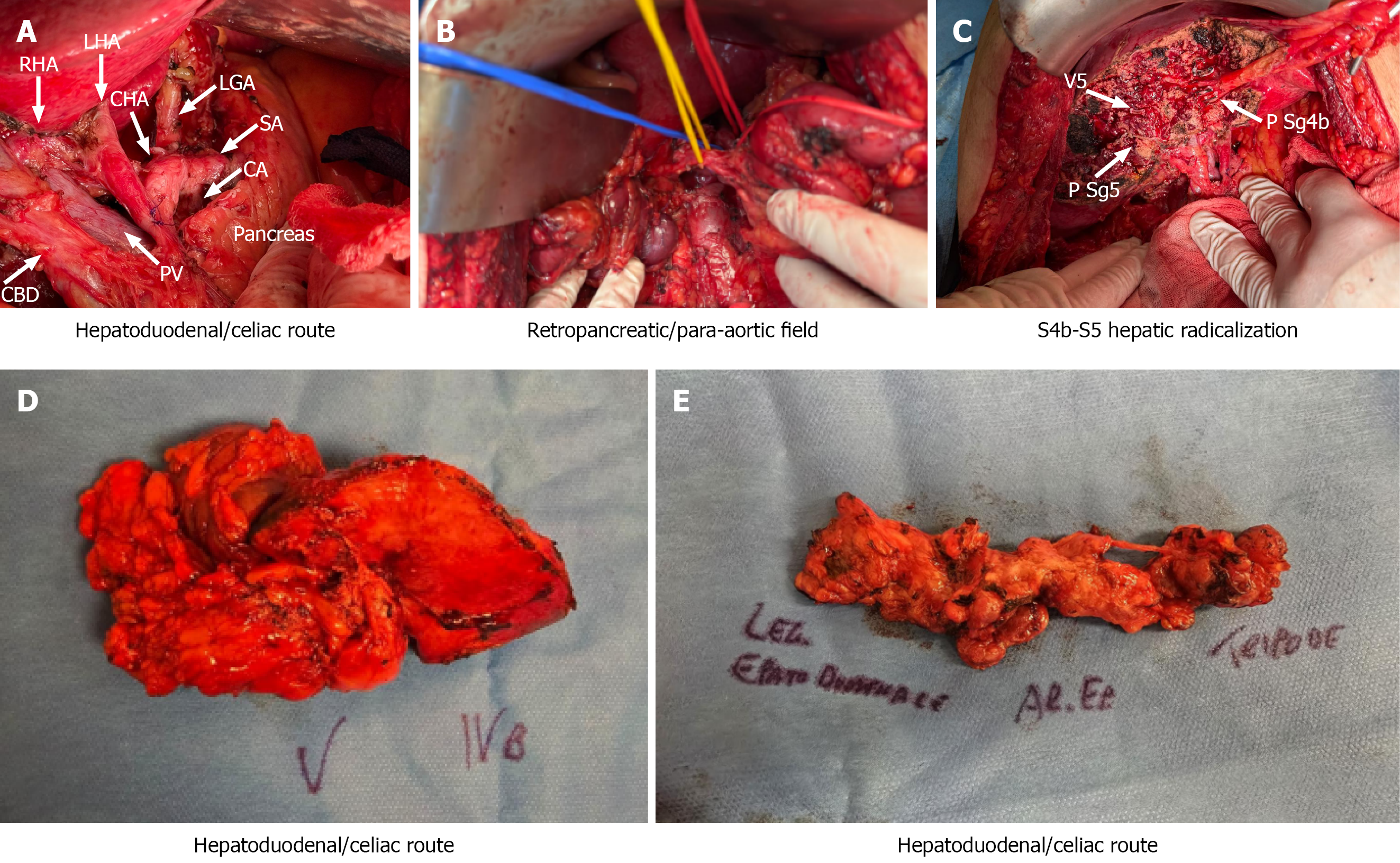
Figure 2 Original intraoperative images illustrating pathway-aware lymphadenectomy and associated radical surgical steps in selected gallbladder cancer.
A: Original anonymized intraoperative image showing hepatoduodenal ligament skeletonization and arterial landmarks during extended hepatobiliary lymphadenectomy after neoadjuvant therapy. The panel supports the discussion of the hepatoduodenal/celiac route; B: Original anonymized intraoperative image illustrating retropancreatic/para-aortic nodal tissue harvesting. The panel supports the conceptual discussion of retropancreatic exposure and convergence-basin assessment, and is not presented as a routine indication; C: Original anonymized intraoperative image of hepatic radicalization involving segments 4b and 5. This panel documents the liver component accompanying selected R0-intent surgery and is not presented as a lymphadenectomy panel; D: Original anonymized image of hepatic radicalization specimen. It is included to show the resection component paired with nodal assessment in selected cases; E: Original anonymized image of en bloc nodal tissue corresponding to stations 12, 8 and 9. This panel supports the concept of station-specific specimen reporting. Original image from the authors’ surgical experience. Reproduced courtesy of the General Surgery Unit, Enna Hospital, with written patient consent and institutional authorization. RHA: Right hepatic artery; LHA: Left hepatic artery; CHA: Common hepatic artery; LGA: Left gastric artery; SA: Splenic artery; CA: Celiac axis.
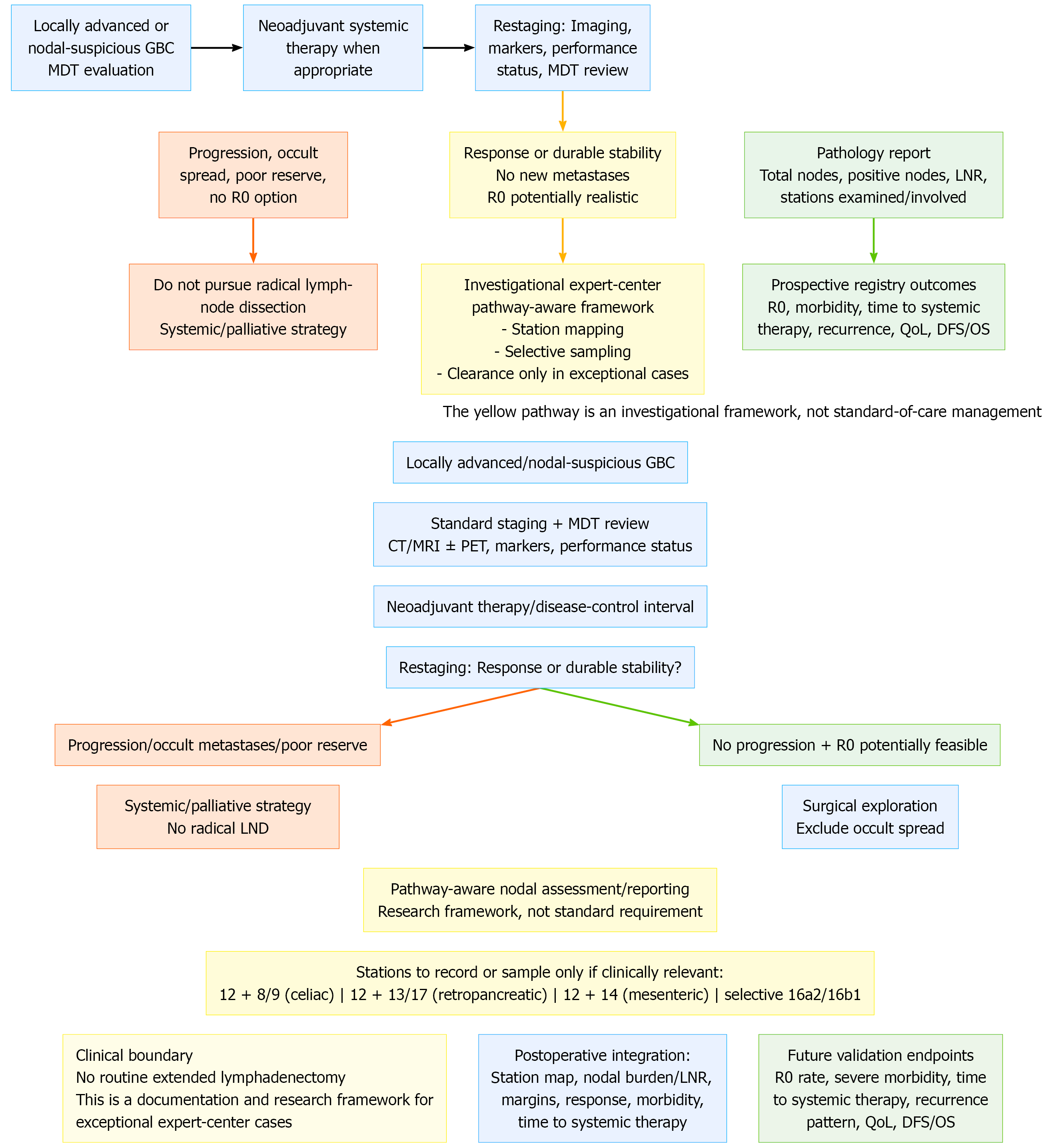
Figure 3 Proposed algorithm for biology-driven, pathway-aware nodal assessment after neoadjuvant selection in locally advanced or nodal-suspicious gallbladder cancer.
The left branch identifies situations in which radical lymph-node dissection should not be pursued. The yellow box marks the authors’ investigational framework rather than standard-of-care management. GBC: Gallbladder cancer; HPB: Hepatopancreatobiliary; LND: Lymph-node dissection; LNR: Lymph-node ratio; MDT: Multidisciplinary team; QoL: Quality of life; DFS: Disease-free survival; OS: Overall survival; CT: Computed tomography; MRI: Magnetic resonance imaging; PET: Positron emission tomography.
- Citation: Cavallaro A, Di Mattia P. Beyond station 16: A pathway-aware framework for gallbladder cancer lymphadenectomy after neoadjuvant therapy. World J Gastrointest Surg 2026; 18(11): 125509
- URL: https://www.wjgnet.com/1948-9366/full/v18/i11/125509.htm
- DOI: https://dx.doi.org/10.4240/wjgs.125509