
















Copyright: ©Author(s) 2026.
World J Gastrointest Surg. Nov 27, 2026; 18(11): 124340
Published online Nov 27, 2026. doi: 10.4240/wjgs.124340
Published online Nov 27, 2026. doi: 10.4240/wjgs.124340
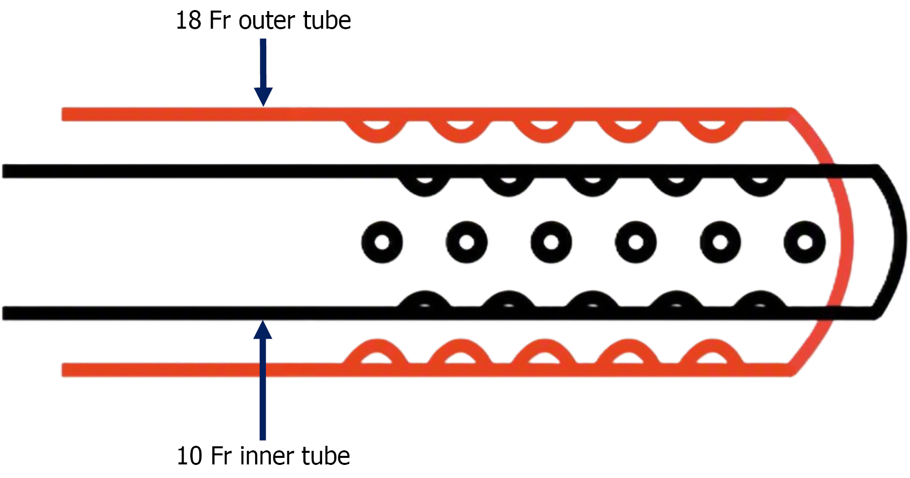
Figure 1 Schematic illustration of the modified vacuum-assisted closure system.
The device consists of a 10-Fr feeding tube inserted through an 18-Fr nasogastric tube. Multiple side holes were created along the distal 3 cm of the feeding tube to allow continuous negative-pressure drainage at the anastomotic site.

Figure 2 Preparation of the modified vacuum-assisted closure device.
A: Components of the modified vacuum-assisted closure system before assembly, including an 18-Fr nasogastric tube and a 10-Fr feeding tube; B: The 10-Fr feeding tube inserted into the lumen of the 18-Fr nasogastric tube, forming the tube-in-tube configuration; C: Creation of multiple side holes along the distal 3 cm of the feeding tube using an 18-gauge needle to facilitate continuous negative-pressure drainage.
- Citation: Ciciboga K, Uprak TK, Ermerak NO. Prophylactic application of modified vacuum-assisted closure therapy in Ivor Lewis esophagectomy: Preliminary results. World J Gastrointest Surg 2026; 18(11): 124340
- URL: https://www.wjgnet.com/1948-9366/full/v18/i11/124340.htm
- DOI: https://dx.doi.org/10.4240/wjgs.124340