
















Copyright: ©Author(s) 2026.
World J Gastrointest Surg. Nov 27, 2026; 18(11): 122899
Published online Nov 27, 2026. doi: 10.4240/wjgs.122899
Published online Nov 27, 2026. doi: 10.4240/wjgs.122899
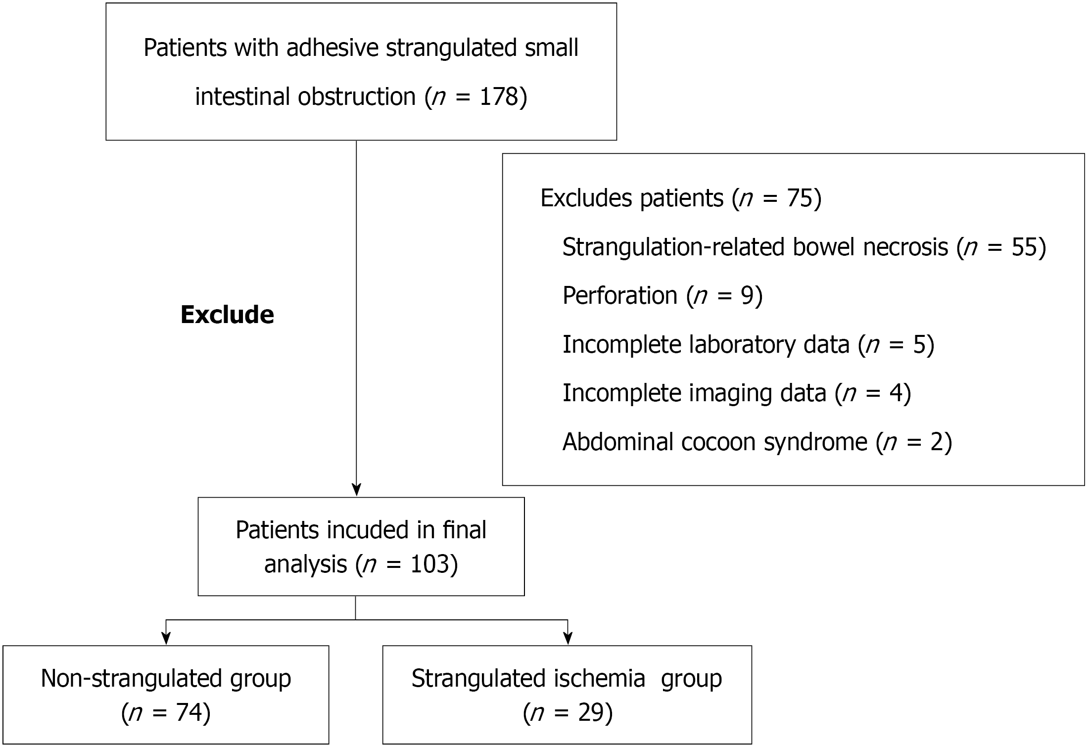
Figure 1
Flowchart of patient selection.
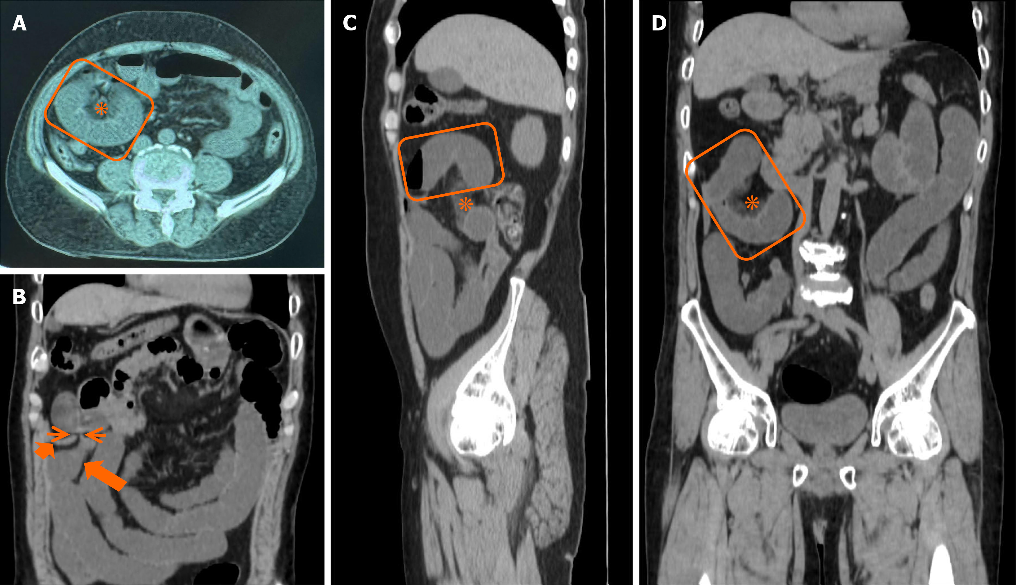
Figure 2 Abdominal computed tomography findings.
A: Axial view = U-/C-shaped bowel loop (orange box) and localized mesenteric haziness (asterisk); B: Coronal view: Closed-loop mechanism (thin orange arrow), collapsed distal bowel (short thick orange arrow), and dilated proximal bowel (long thick orange arrow); C: Sagittal view = U-/C-shaped bowel loop (orange box) and mesenteric haziness (asterisk); D: Coronal view = U-/C-shaped bowel loop (orange box) and mesenteric haziness (asterisk).
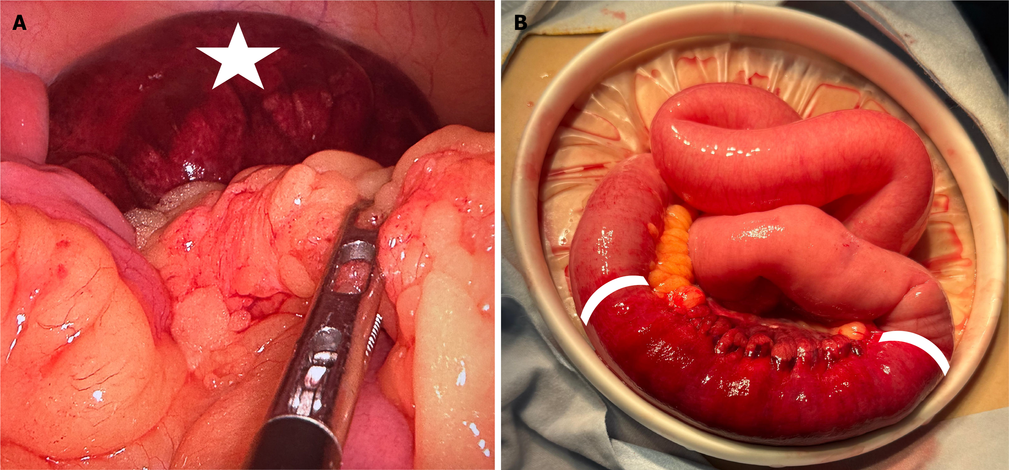
Figure 3 Intraoperative findings.
A: The greater omentum adheres to the mesentery of the ascending colon, forming a defect (forceps). A herniated small bowel segment appears dark purple-red (white star); B: After release of strangulation, bowel color improves (white bracket) and peristalsis returns. No bowel resection was performed.
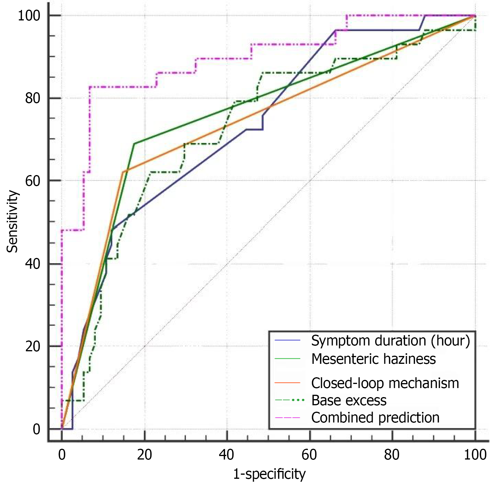
Figure 4 Receiver operating characteristic curves of combined model.
Curves represent: Symptom duration [≤ 20 hours, area under the curve (AUC) = 0.735], mesenteric haziness (AUC = 0.757), closed-loop obstruction (AUC = 0.736), base excess (≤ -3, AUC = 0.727), and the combined model (AUC = 0.897). The combined model shows the highest area under the curve.
- Citation: Dong LC, Zhang JD, Hsieh YF, Xu SC. Early identification of reversible strangulating ischemia in adhesive small bowel obstruction using non-contrast computed tomography and routine laboratory parameters. World J Gastrointest Surg 2026; 18(11): 122899
- URL: https://www.wjgnet.com/1948-9366/full/v18/i11/122899.htm
- DOI: https://dx.doi.org/10.4240/wjgs.122899