Copyright
©The Author(s) 2025.
World J Gastrointest Surg. Jun 27, 2025; 17(6): 106531
Published online Jun 27, 2025. doi: 10.4240/wjgs.v17.i6.106531
Published online Jun 27, 2025. doi: 10.4240/wjgs.v17.i6.106531
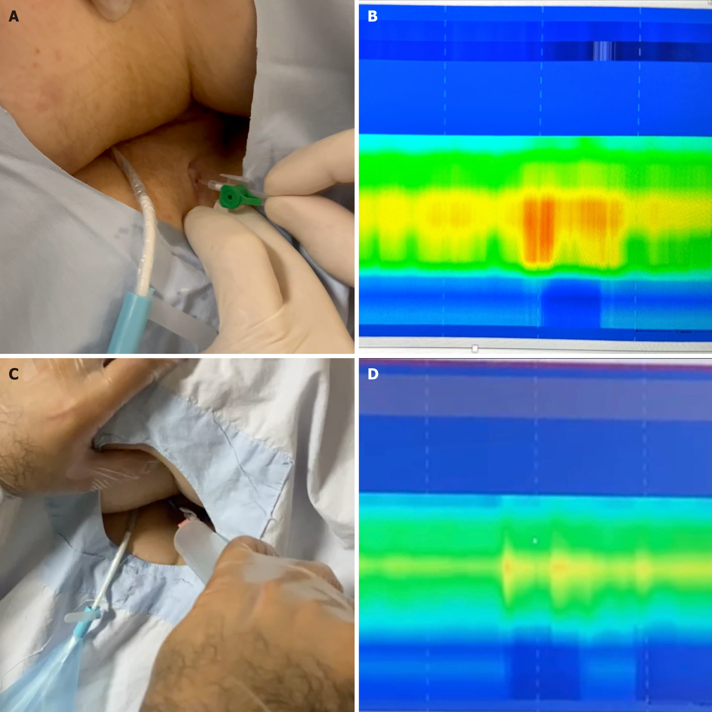
Figure 1 Comparison of the preoperative high-resolution manometry (when the internal orifice of the fistula is at the high-pressure zone) and postoperative high-resolution manometry (after the distalization of the internal orifice to the low-pressure zone).
A: Air injection through the fistula for anal manometry: An anal manometry catheter is placed in the anal canal and air is injected into the external orifice of the fistula with an angiocath; B: Preoperative manometry: Preoperative measurement of the high anal canal pressure, with orange areas indicating high pressure zones; C: Air injection to assess post-distalization pressure: Injection of air from the anal canal with an angiocath to measure anal pressure after distalization; D: Post-distalization manometry showing reduced pressure: After distalization, high-resolution manometry shows lower anal pressure, now represented by yellow areas instead of orange in the preoperative state.
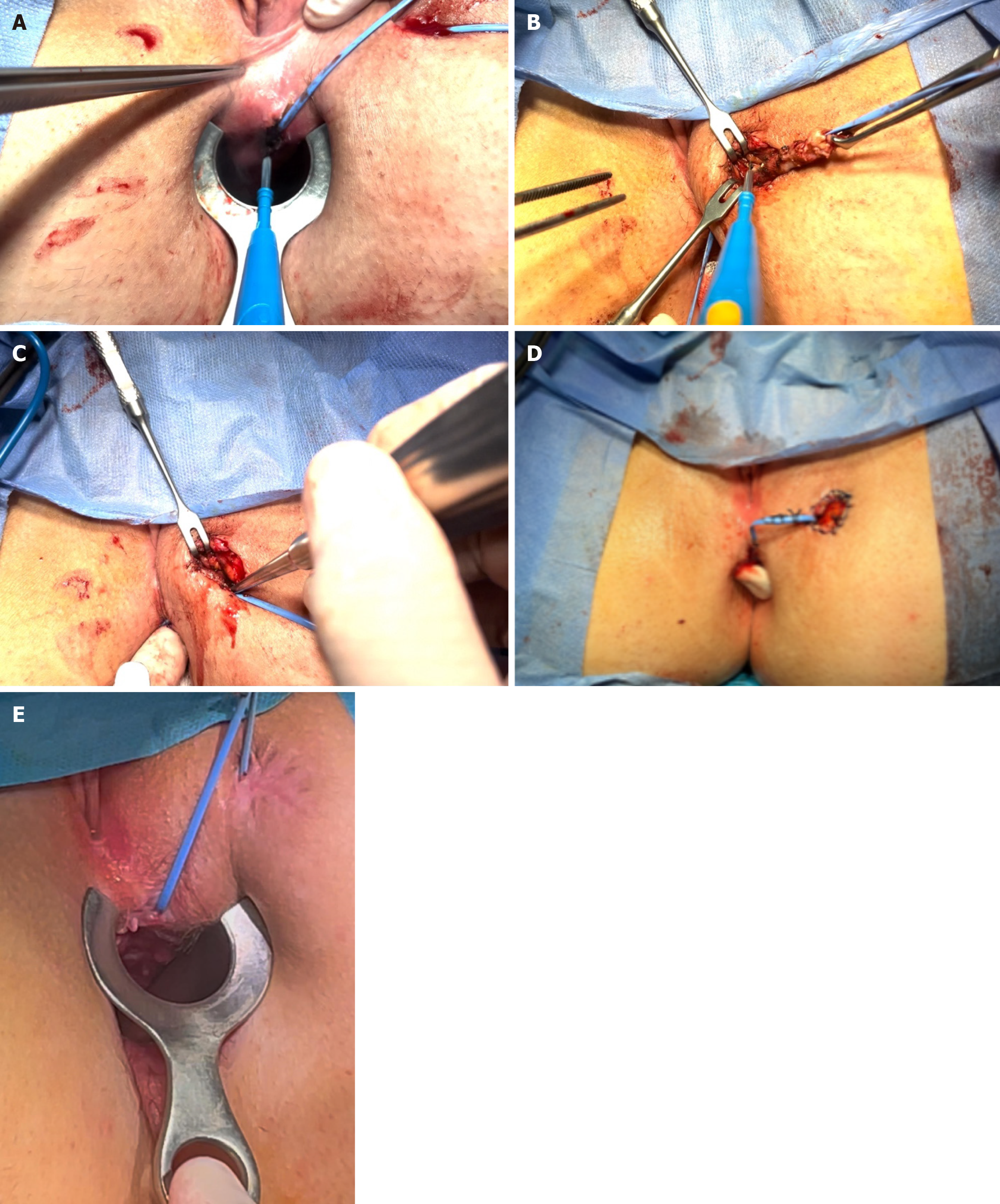
Figure 2 Key steps of the modified fistulotomy technique.
A: Distalization of the internal orifice achieved by incising the anoderm and partially cutting the internal sphincter up to the intersphincteric groove; B: Excision of the fistula tract up to the external sphincter muscle; C: Curettage of the remaining fistula tract; D: Distalization of the internal opening; E: Internal and external openings without signs of inflammation at postoperative month 3.
- Citation: Eray İC, Yavuz B, Aydin I, Gumus S, Topal U, Dalci K. Modified fistulotomy with internal orifice distalization for optimized perianal fistula management: Pressure zone transition. World J Gastrointest Surg 2025; 17(6): 106531
- URL: https://www.wjgnet.com/1948-9366/full/v17/i6/106531.htm
- DOI: https://dx.doi.org/10.4240/wjgs.v17.i6.106531


