Copyright
©The Author(s) 2024.
World J Gastrointest Surg. Apr 27, 2024; 16(4): 1043-1054
Published online Apr 27, 2024. doi: 10.4240/wjgs.v16.i4.1043
Published online Apr 27, 2024. doi: 10.4240/wjgs.v16.i4.1043
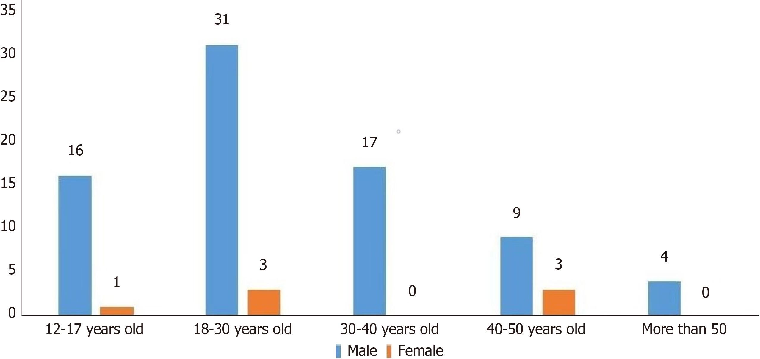
Figure 1 Age and sex proportion of 84 Meckel’s diverticulum hemorrhage patients.
Incidence was significantly higher in men than in women (P < 0.01).
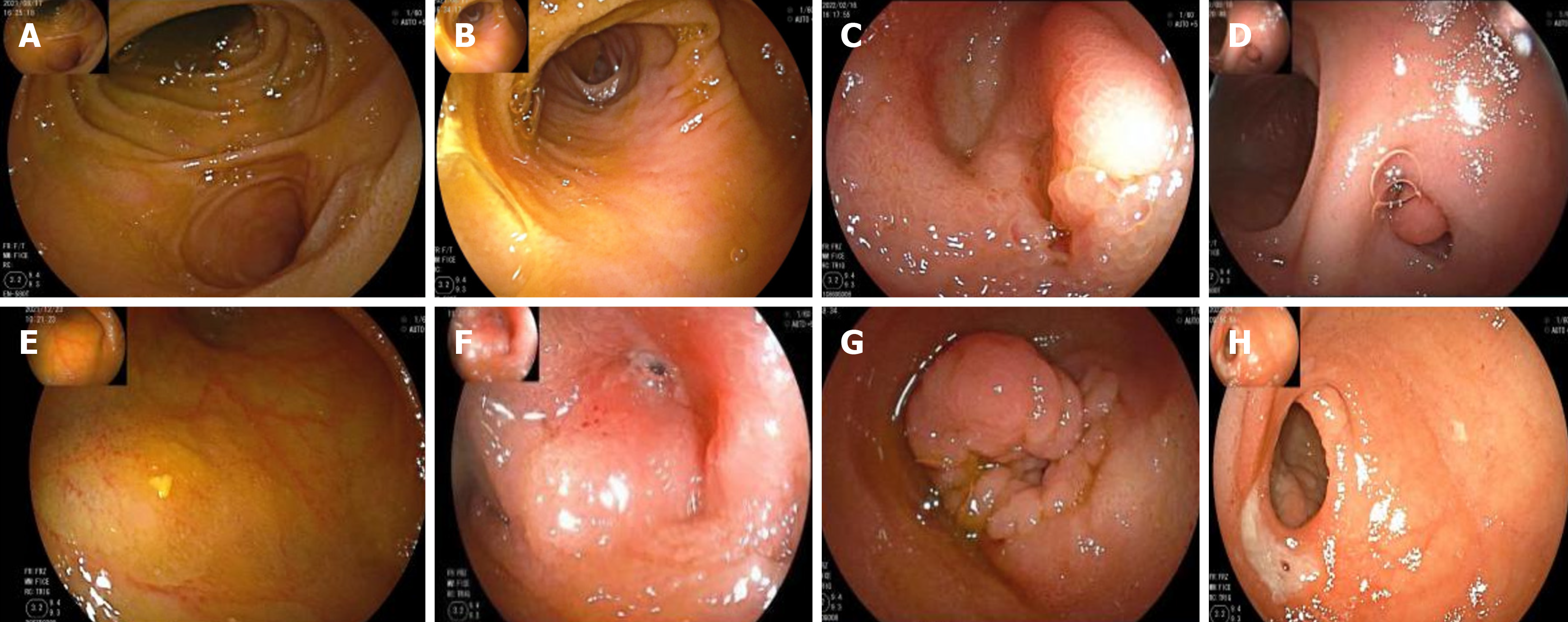
Figure 2 Appearance of Meckel’s diverticulum by double-balloon enteroscopy.
A: Mucosal ridge; B: Blind end of diverticulum; C: Inflammation in the diverticulum; D: Diverticulum varus; E: Ulceration in diverticulum; F: Blind ulcer stenosis; G: Diverticulum varus hyperplasia; H: Edge ulcer of the diverticulum.
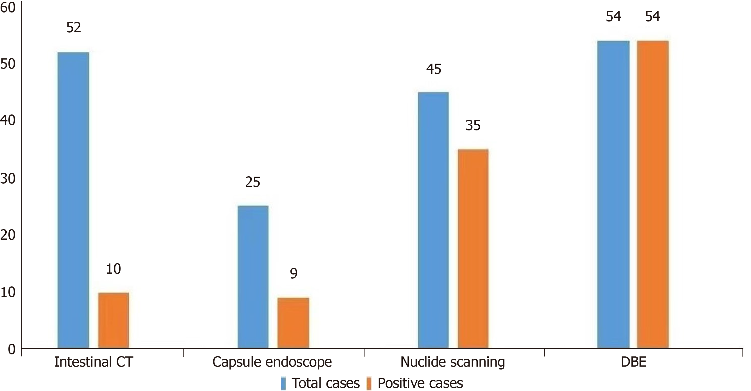
Figure 3 Diagnosis of Meckel’s diverticulum complicated by hemorrhage.
CT: Computed tomography; DBE: Double-balloon enteroscopy.
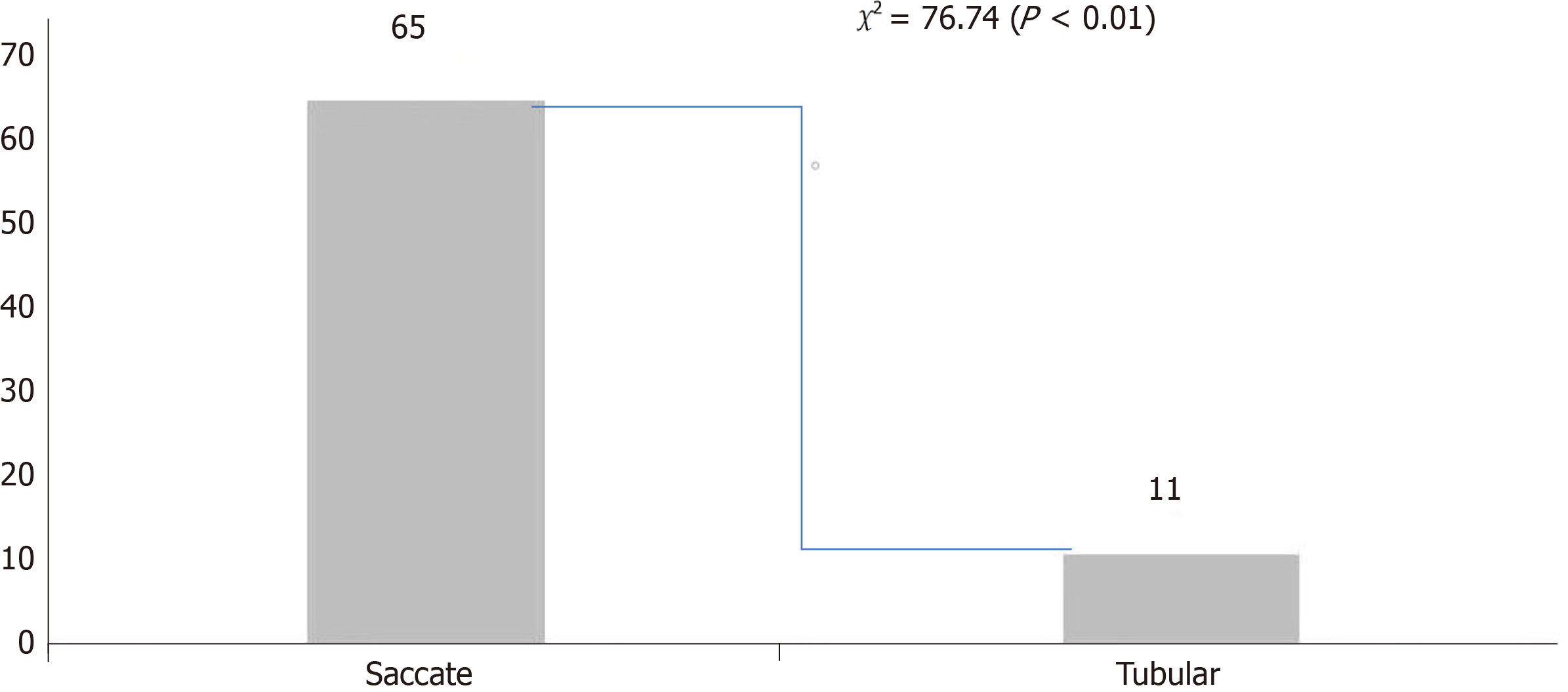
Figure 4 Morphological analysis of Meckel’s.
diverticulum.P < 0.01 compared with tubular diverticulum, the incidence of cystic Meckel’s diverticulum (MD) is higher in MD with hemorrhage.
- Citation: He T, Yang C, Wang J, Zhong JS, Li AH, Yin YJ, Luo LL, Rao CM, Mao NF, Guo Q, Zuo Z, Zhang W, Wan P. Single-center retrospective study of the diagnostic value of double-balloon enteroscopy in Meckel’s diverticulum with bleeding. World J Gastrointest Surg 2024; 16(4): 1043-1054
- URL: https://www.wjgnet.com/1948-9366/full/v16/i4/1043.htm
- DOI: https://dx.doi.org/10.4240/wjgs.v16.i4.1043


