Copyright
©The Author(s) 2024.
World J Gastrointest Surg. Apr 27, 2024; 16(4): 1030-1042
Published online Apr 27, 2024. doi: 10.4240/wjgs.v16.i4.1030
Published online Apr 27, 2024. doi: 10.4240/wjgs.v16.i4.1030
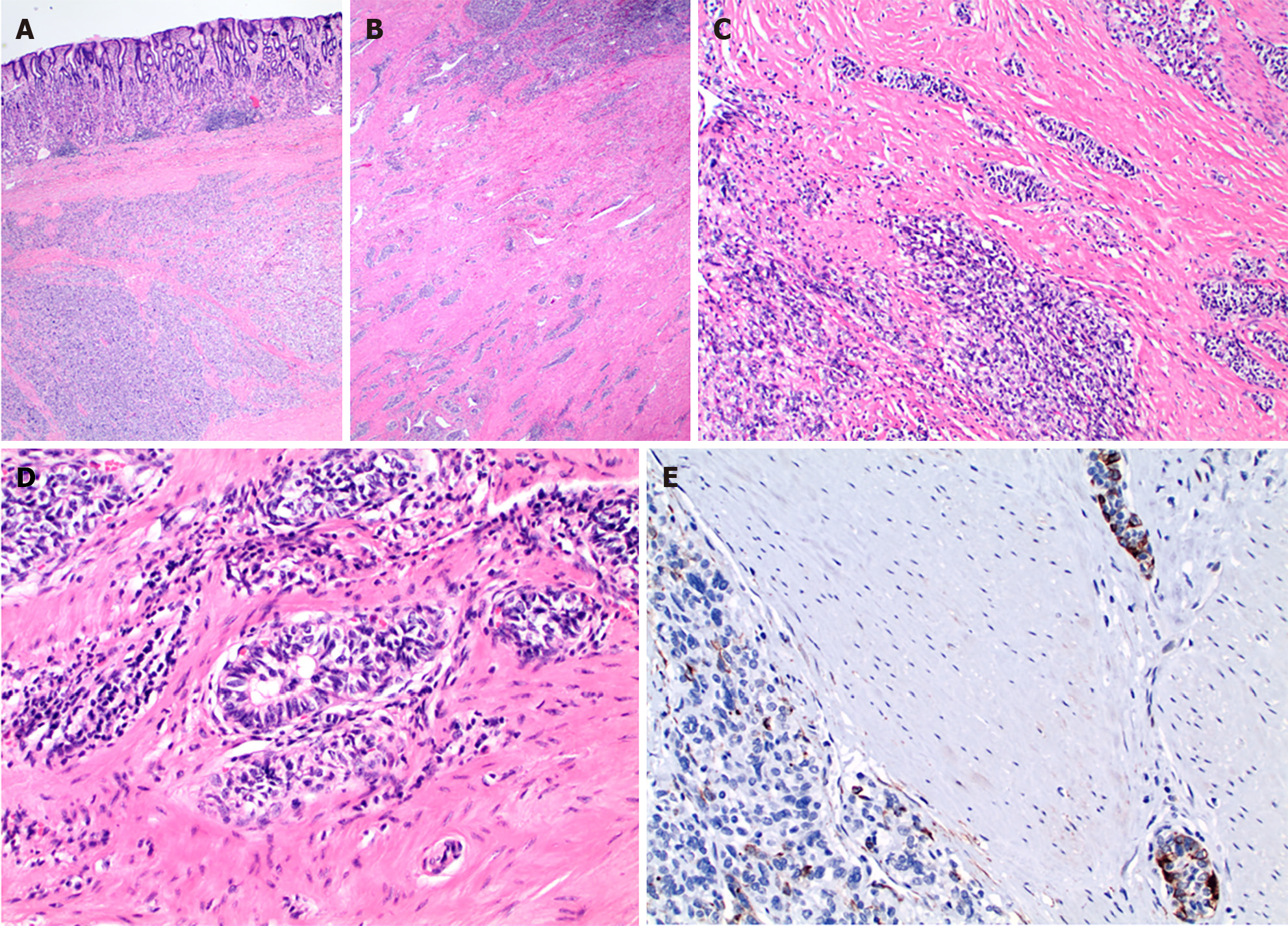
Figure 1 Gastroblastoma in a 19-year-old patient.
A and B: The tumor involves the muscularis propria of the antrum (20 ×); C: The tumor shows a biphasic histology with spindle stromal component more prominent in left lower of the image (40 ×); D: The high-magnification image shows the epithelial cells with glandular formation (400 ×); E: Cytoketatin staining (AE1/3) labels the epithelial cells but not the mesenchymal component (200 ×).
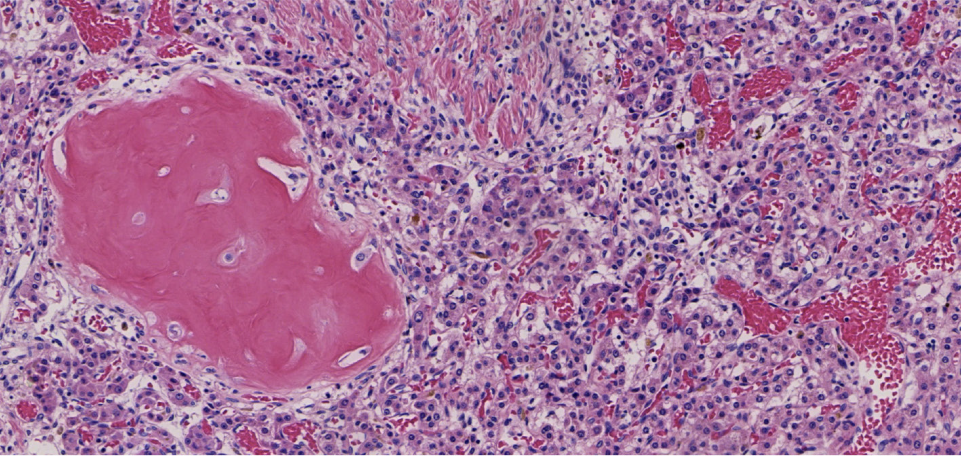
Figure 2 Mixed epithelial mesenchymal hepatoblastoma in an adult patient, showing a heterogeneous mix of epithelial and mesenchymal components.
Well differentiated fetal-type epithelial component is characterized by their eosinophilic cytoplasm and cords or nests-like growth reminiscent of normal liver architecture. The mesenchymal part shows heterologous osteoid elements (100 ×).
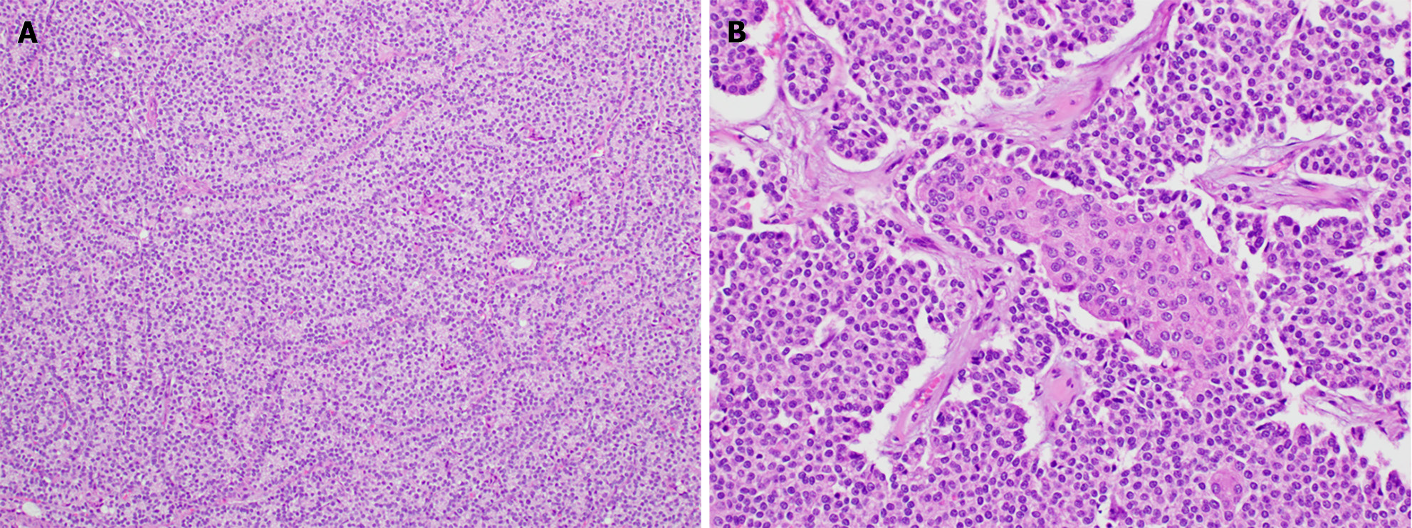
Figure 3 Pancreatoblastoma in the pancreatic head of a 42-year-old male patient.
A: The tumor is composed of acinar and ribbonlike morphology composed of relatively monotonous cells with basophilic nuclei, mimicking a pancreatic endocrine tumor (100 ×); B: In a few foci, squamoid nests, consisting of circumscribed whorled nests of polygonal cells with abundant eosinophilic cytoplasm and a squamous appearance, are present (200 ×).
- Citation: Liu Y, El Jabbour T, Somma J, Nakanishi Y, Ligato S, Lee H, Fu ZY. Blastomas of the digestive system in adults: A review. World J Gastrointest Surg 2024; 16(4): 1030-1042
- URL: https://www.wjgnet.com/1948-9366/full/v16/i4/1030.htm
- DOI: https://dx.doi.org/10.4240/wjgs.v16.i4.1030


