Copyright
©The Author(s) 2024.
World J Gastrointest Surg. Jan 27, 2024; 16(1): 113-123
Published online Jan 27, 2024. doi: 10.4240/wjgs.v16.i1.113
Published online Jan 27, 2024. doi: 10.4240/wjgs.v16.i1.113
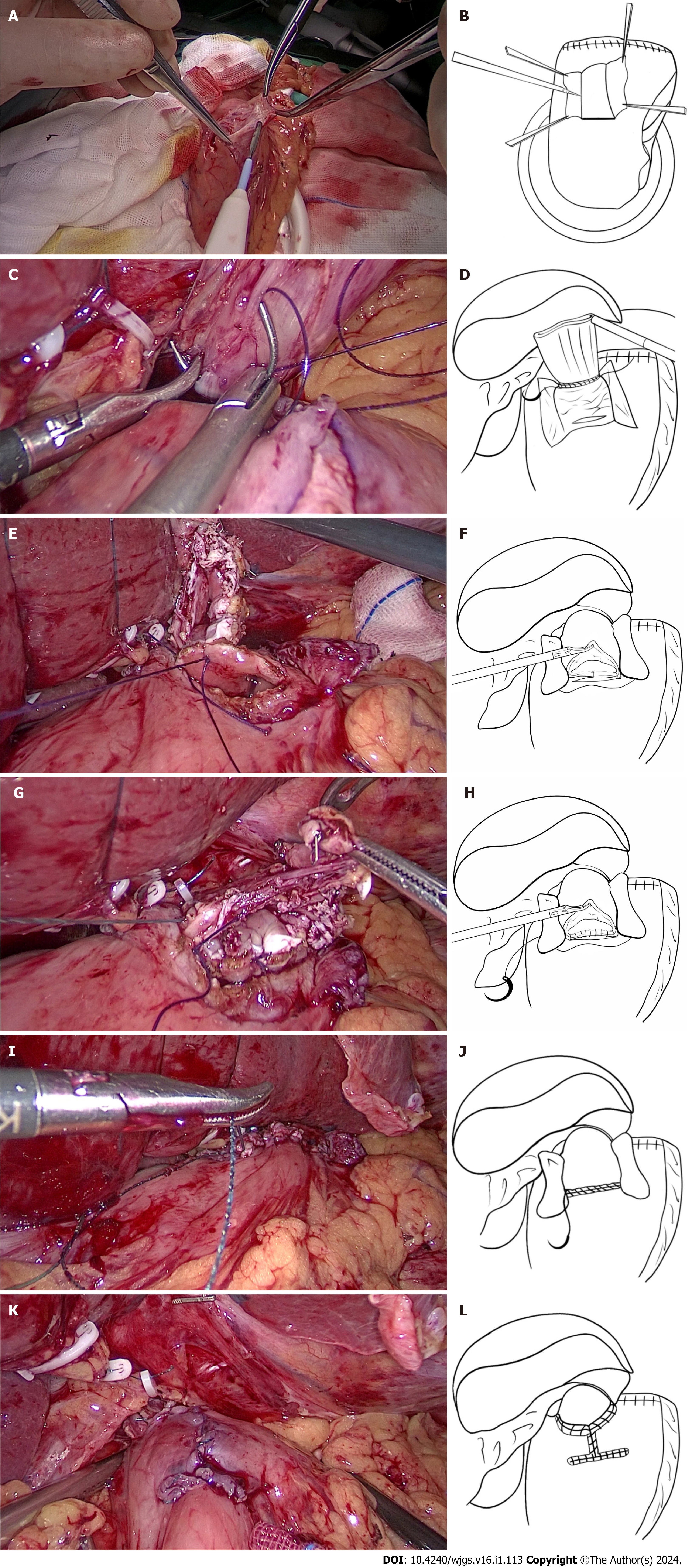
Figure 1 Laparoscopic proximal gastrectomy and modified Kamikawa anastomosis were performed.
A and B: The surgeon used an electrotome to completely dissect the submucosal layer from the mucosa to create the seromuscular flap; C and D: The region marked with gentian violet at the lower esophagus and the superior border of the seromuscular flap was continuously sutured and fixed with a 3-0 laparoscopic line with an arc of 5/8; E and F: The posterior wall of the esophageal stump opening and the superior border of the anastomotic stoma were first fixed by placing two interrupted sutures on the left and in the middle; G and H: A 3-0 barbed suture was used for continuous suturing of the full thickness of the posterior wall of the esophageal stump opening and the gastric mucosa and submucosa at the superior border of the anastomotic stoma, starting from the left part and proceeding to the right edge; I and J: A second 3-0 barbed continuous suture was used to suture the full thickness of the anterior wall of the esophageal stump opening and the stomach at the inferior border of the anastomotic stoma, starting from the left part and proceeding to the right edge; K and L: The blood supply in the seromuscular flap was observed.
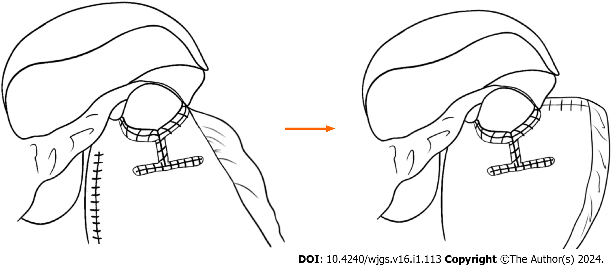
Figure 2 Our modifications compared with traditional Kamikawa anastomosis.
- Citation: Wu CY, Lin JA, Ye K. Clinical efficacy of modified Kamikawa anastomosis in patients with laparoscopic proximal gastrectomy. World J Gastrointest Surg 2024; 16(1): 113-123
- URL: https://www.wjgnet.com/1948-9366/full/v16/i1/113.htm
- DOI: https://dx.doi.org/10.4240/wjgs.v16.i1.113


