Copyright
©The Author(s) 2021.
World J Gastrointest Surg. Aug 27, 2021; 13(8): 788-795
Published online Aug 27, 2021. doi: 10.4240/wjgs.v13.i8.788
Published online Aug 27, 2021. doi: 10.4240/wjgs.v13.i8.788
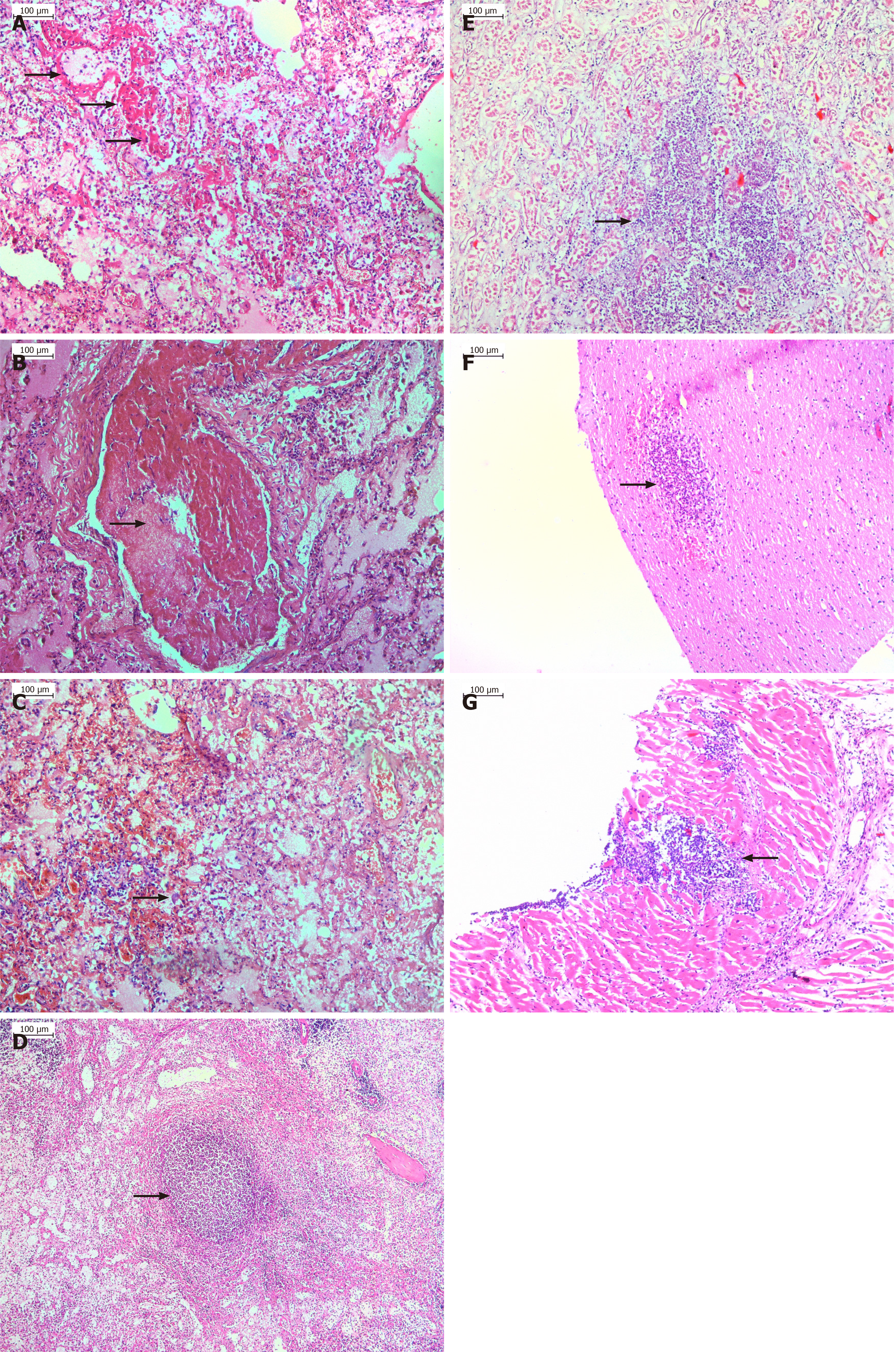
Figure 1 Histology examination.
A: Hyaline membranes (marked with an arrow) covering the alveolar walls in a case of septic acute respiratory distress syndrome originating from gangrenous appendicitis in coronavirus disease-2019 patient; B: Microthrombosis resulted in almost complete obstruction of the pulmonary vessel; C: Diffuse alveolar damage with interalveolar hemorrhages and inflammatory cell infiltration, as well as type II hyperplastic pneumocyte (marked with an arrow); D-G: Septicopyemic abscess in the spleen (D, marked with an arrow), kidney (E, marked with an arrow), brain (F, marked with an arrow) and myocardium (G, marked with an arrow); E: Acute tubular necrosis found.
- Citation: Gulinac M, Novakov IP, Antovic S, Velikova T. Surgical complications in COVID-19 patients in the setting of moderate to severe disease. World J Gastrointest Surg 2021; 13(8): 788-795
- URL: https://www.wjgnet.com/1948-9366/full/v13/i8/788.htm
- DOI: https://dx.doi.org/10.4240/wjgs.v13.i8.788


