Copyright
©The Author(s) 2020.
World J Gastrointest Surg. Jun 27, 2020; 12(6): 277-286
Published online Jun 27, 2020. doi: 10.4240/wjgs.v12.i6.277
Published online Jun 27, 2020. doi: 10.4240/wjgs.v12.i6.277
Figure 1 Group 2 lymph nodes and group 1 lymph nodes for distal gastrectomy.
A: Group 2 lymph nodes are suprapancreatic nodes and were harvested using the conventional method; B: Group 1 nodes are perigastric nodes embedded in adipose tissue and were harvested using the fat-dissociation method.
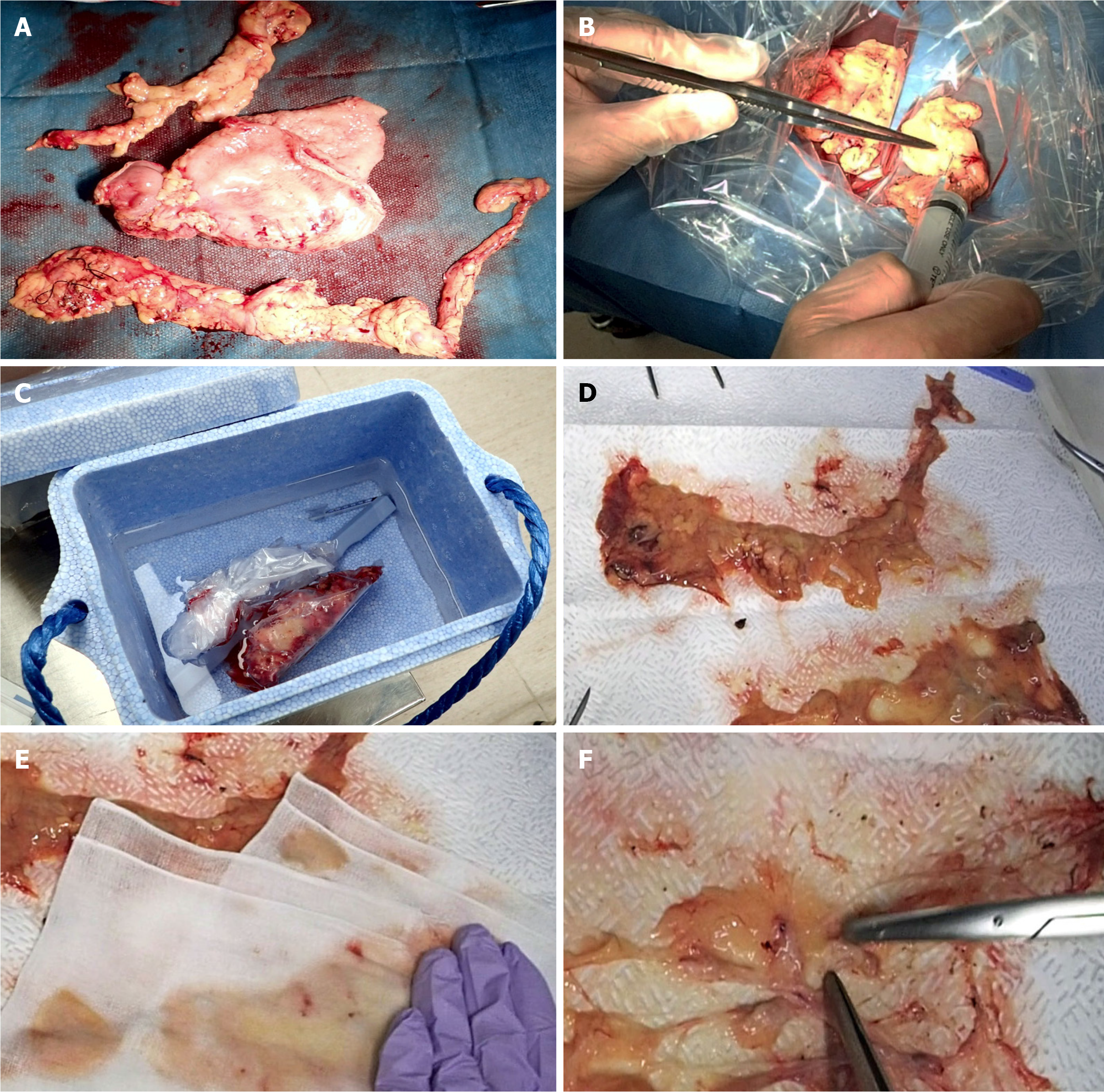
Figure 2 Fat-dissociation method for gastric cancer.
A: The adipose tissue, including group 1 nodes, was detached en bloc from the stomach; B: The fat dissolving reagent (Imofully®) was injected into the tissue using a syringe; C: The tissue was put into a nylon bag and was rubbed manually by fingers from the outside. Then, the nylon bag was soaked in hot water at 42 °C and incubated for 1 h; D: The tissue was carefully spread on a water-absorbing paper (Kim Towel®) on the table; E: A gauze was added, and pressure was applied to remove the dissolved fat; F: After the fat droplets had been sufficiently absorbed, the lymph nodes were identified, picked up with scissors, and mapped.
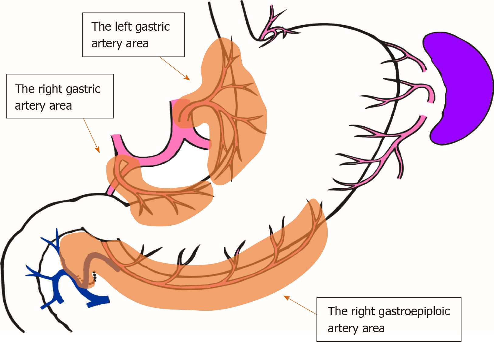
Figure 3 The lymphatic compartments used in this study are shown.
- Citation: Kinami S, Ohnishi T, Nakamura N, Jiang ZY, Miyata T, Fujita H, Takamura H, Ueda N, Kosaka T. Efficacy of the fat-dissociation method for nodal harvesting in gastric cancer. World J Gastrointest Surg 2020; 12(6): 277-286
- URL: https://www.wjgnet.com/1948-9366/full/v12/i6/277.htm
- DOI: https://dx.doi.org/10.4240/wjgs.v12.i6.277


