Copyright
©The Author(s) 2020.
World J Gastrointest Surg. Dec 27, 2020; 12(12): 555-563
Published online Dec 27, 2020. doi: 10.4240/wjgs.v12.i12.555
Published online Dec 27, 2020. doi: 10.4240/wjgs.v12.i12.555
Figure 1 Initial endoscopic and pathology examination.
A: Esophagogastroduodenoscopy revealed several ulcers in the angle and antrum of the stomach; B: Pathology suggested low grade adenocarcinoma (× 100). Immunohistochemistry revealed HER-2(-); C: The biopsy pathology of the rib suggested metastatic adenocarcinoma (× 100). Immunohistochemistry showed CK7(-), CK20(+), CDX2(+), TTF-1(-), NapsinA (-), and TG (-).
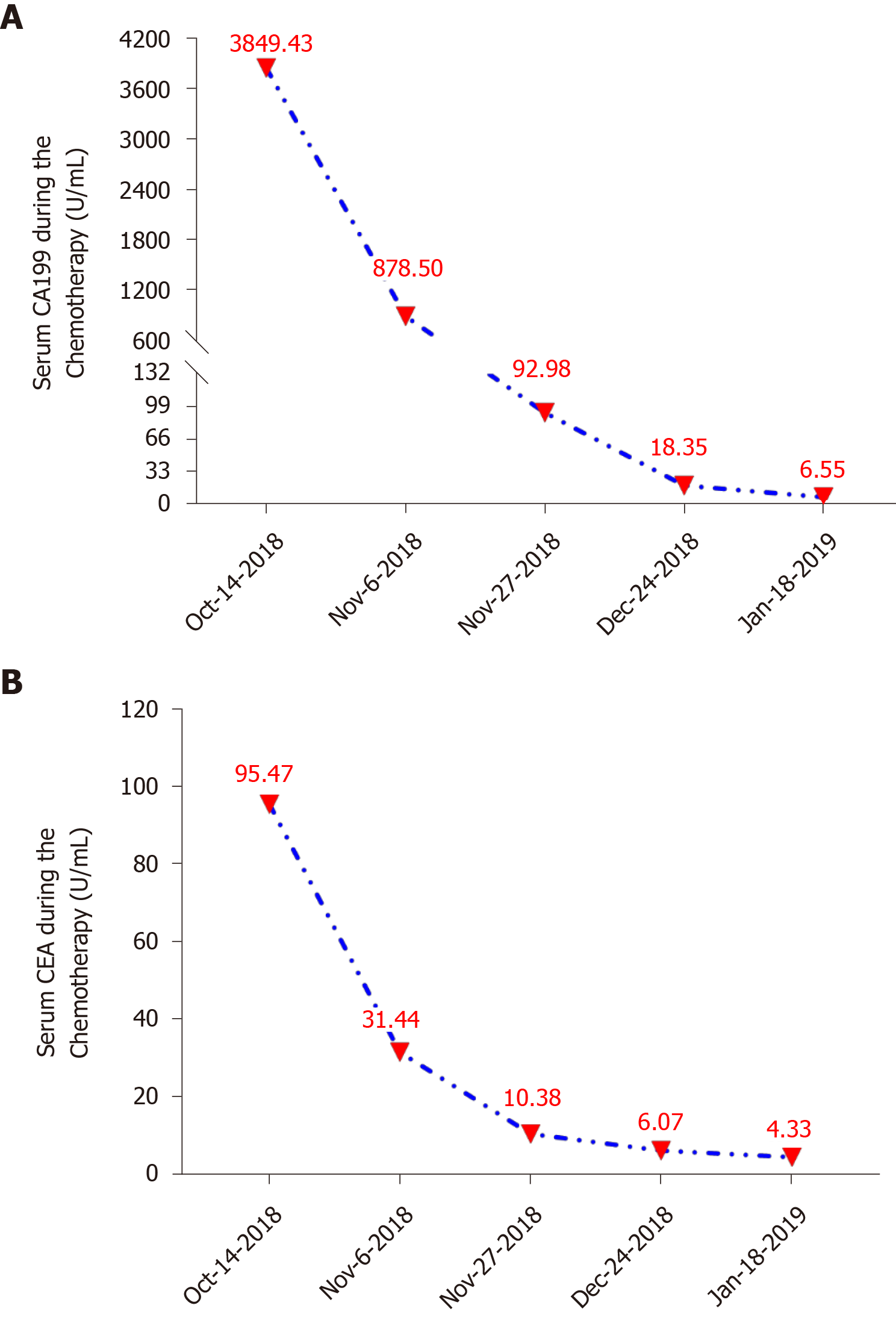
Figure 2 Changes in the levels of tumor biomarkers during adjuvant chemotherapy.
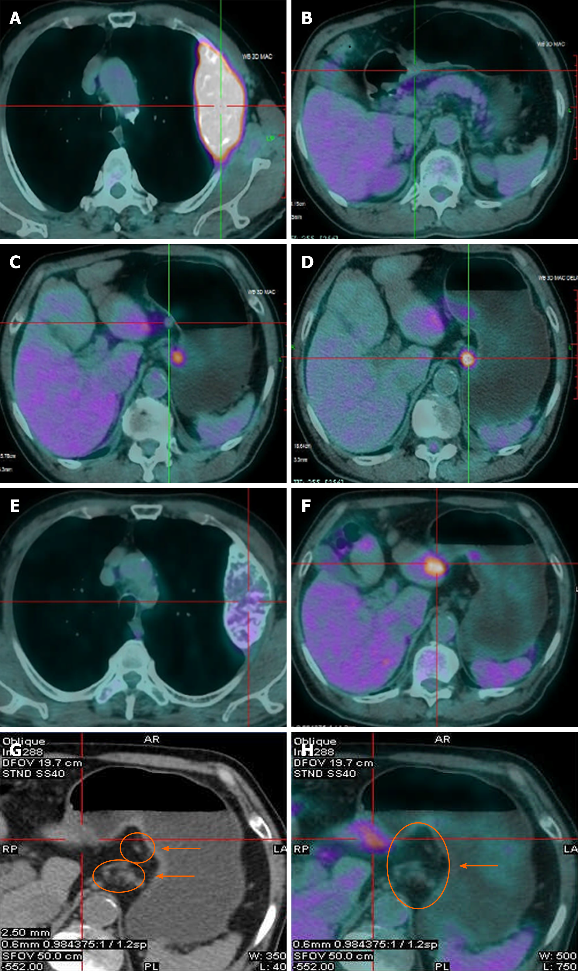
Figure 3 Positron emission tomography/chest computed tomography scanning presentation at different times.
A-D: Baseline lesions. A: Rib bone metastasis, SUVmax = 8.08; B: Gastric tumor, SUVmax = 3.65; C: Metastatic lymph nodes 1, SUVmax = 6.5; D: Metastatic lymph nodes 2, SUVmax = 3.2. E-H: Changes in the target lesions after five courses of chemotherapy. E: Rib metastasis, SUVmax = 2.65; F: Gastric tumor, SUVmax = 3.58; G and H: Metastatic lymph nodes, SUVmax = 0.
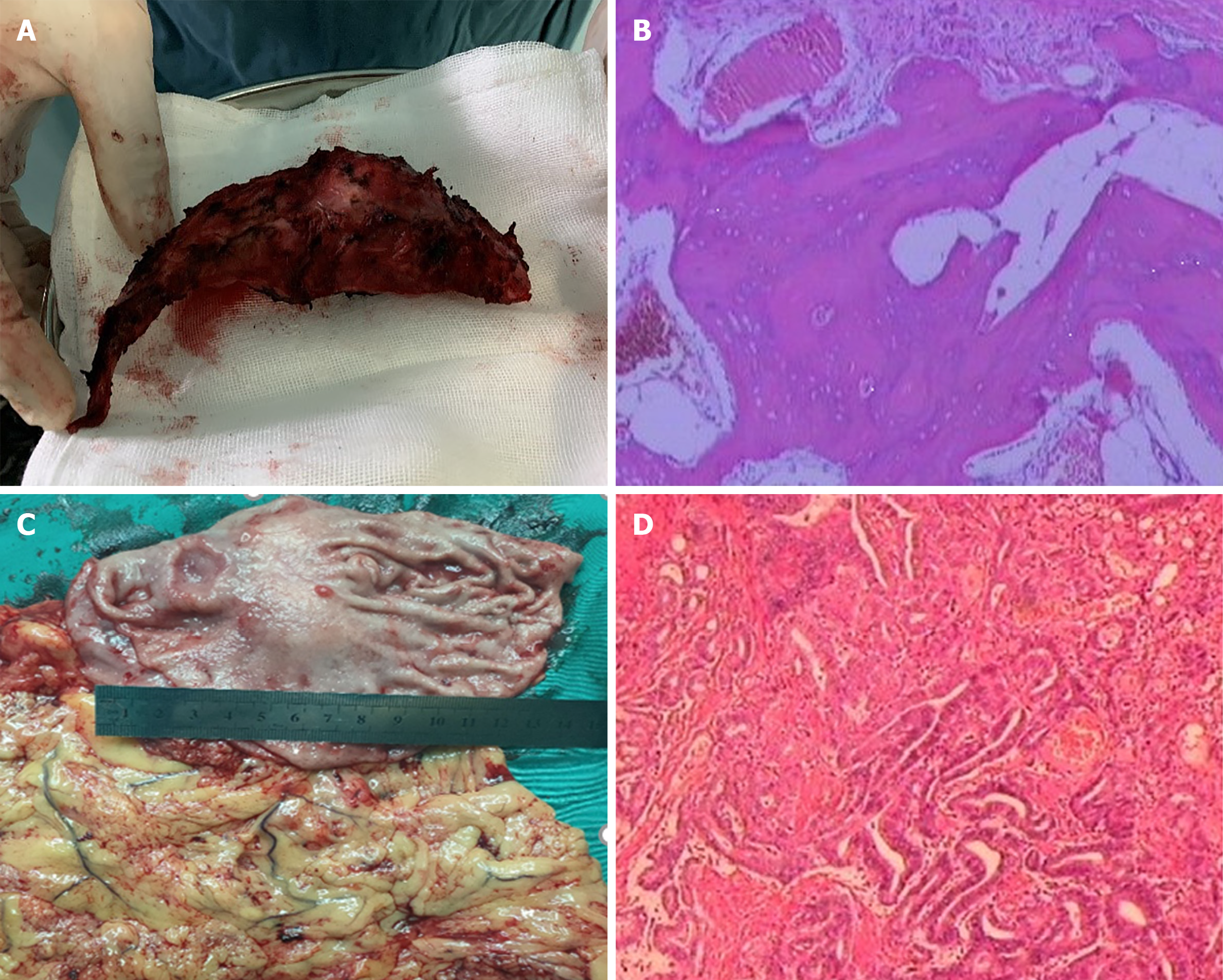
Figure 4 Postoperative specimens and pathologies.
A: Left 3rd rib metastasis; B: Pathology of metastasis, with complete regression and no metastatic tumor left (tumor regression grade classification: 0, × 100); C: Gastric cancer; D: Pathology of ulcerative gastric moderately differentiated adenocarcinoma that infiltrated into the submucosa and adjacent to the superficial muscle layer locally, low-grade intraepithelial neoplasia of some glands in the surrounding mucosa, and tumor stroma infiltrated with a small amount of inflammatory cells, with 1/36 lymph node metastasis (partial response, tumor regression grade classification: 2, × 100). Immunohistochemistry revealed CD34(+), blood vessels(+), CDX2(+), CgA(-), CK20(-), CK7(+), CKpan(+), CyclinD1(+), EGFR(+), Ki-67 (+75%), P53(+), Survivin(+), Syn(-), CerbB-2(2+), MLH1(+), MSH2(+), MSH6(+), and PMS2(+).
- Citation: Zhang Y, Zhang ZX, Lu ZX, Liu F, Hu GY, Tao F, Ye MF. Individualized treatment for gastric cancer with rib metastasis: A case report. World J Gastrointest Surg 2020; 12(12): 555-563
- URL: https://www.wjgnet.com/1948-9366/full/v12/i12/555.htm
- DOI: https://dx.doi.org/10.4240/wjgs.v12.i12.555


