Copyright
©The Author(s) 2019.
World J Gastrointest Surg. Jan 27, 2019; 11(1): 27-33
Published online Jan 27, 2019. doi: 10.4240/wjgs.v11.i1.27
Published online Jan 27, 2019. doi: 10.4240/wjgs.v11.i1.27
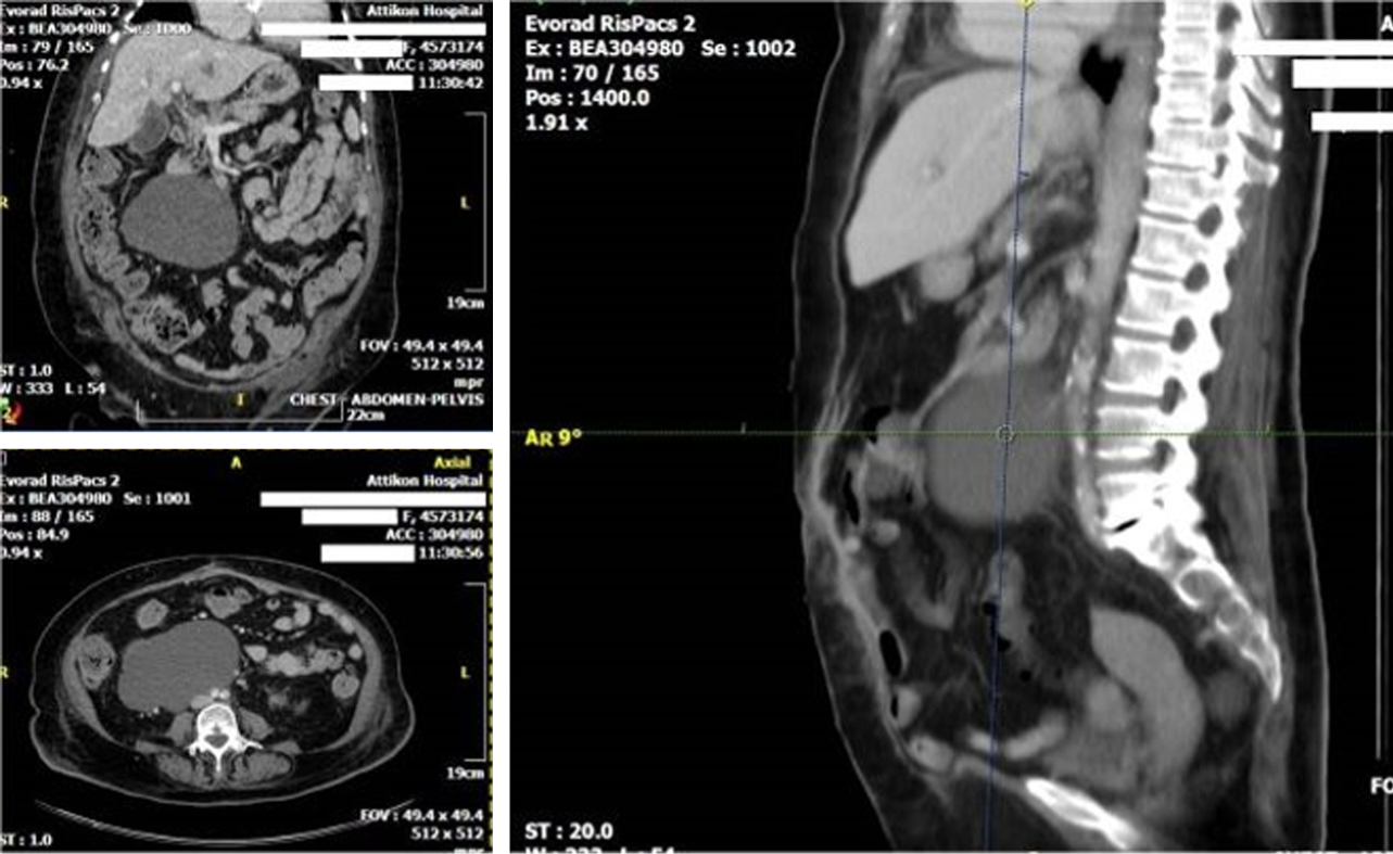
Figure 1 Computerized tomography depiction of a concentric, sharply marginated retro-peritoneal synovial sarcoma.
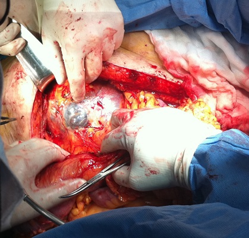
Figure 2 Primary retroperitoneal synovial sarcoma with solid and cystic areas and residues of haemorrhage.
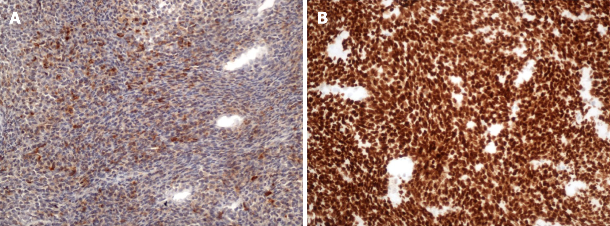
Figure 3 Immunohistochemical stain.
A: Immunohistochemical stain for TLE-1 with parallel strong nuclear expression for the marker (200× magnification); B: Immunohistochemical stain for cytokeratin AE1/AE3 with diffuse expression for the marker (200× magnification).
- Citation: Mastoraki A, Schizas D, Papanikolaou IS, Bagias G, Machairas N, Agrogiannis G, Liakakos T, Arkadopoulos N. Management of primary retroperitoneal synovial sarcoma: A case report and review of literature. World J Gastrointest Surg 2019; 11(1): 27-33
- URL: https://www.wjgnet.com/1948-9366/full/v11/i1/27.htm
- DOI: https://dx.doi.org/10.4240/wjgs.v11.i1.27


