Copyright
©The Author(s) 2018.
World Journal of Gastrointestinal Surgery. Aug 27, 2018; 10(5): 49-56
Published online Aug 27, 2018. doi: 10.4240/wjgs.v10.i5.49
Published online Aug 27, 2018. doi: 10.4240/wjgs.v10.i5.49
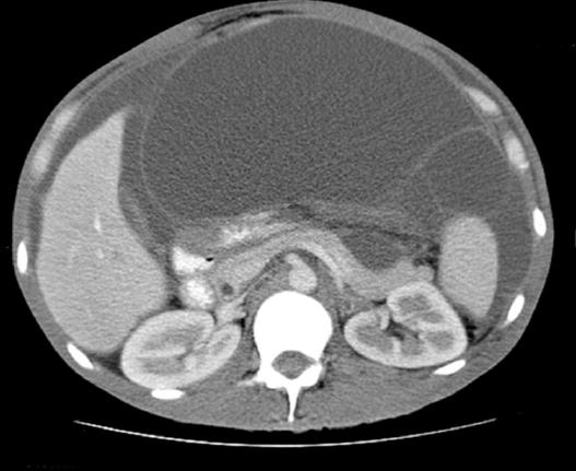
Figure 1 Mucinous ascites on computed tomography[24].
This computed tomography scan demonstrates mucinous ascites. We also see characteristic findings of pseudomyxoma peritonei: Scalloping of the liver and spleen.
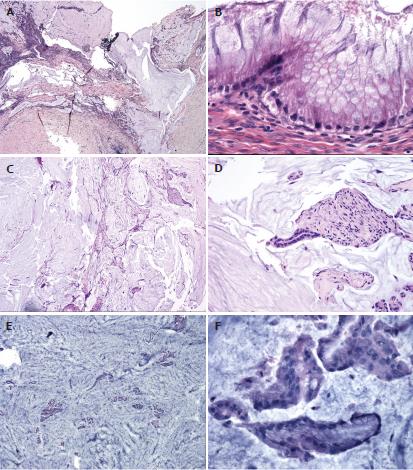
Figure 2 Histological classification of pseudomyxoma peritonei[25].
This figure highlights some of the characteristic findings in the different histological types of pseudomyxoma peritonei. A, B: Disseminated peritoneal adenomucinous (DPAM) is demonstrated in (A and B) with paucicellular mucin pools (A) and scant strips of low-grade neoplastic epithelium (B); C, D: Peritoneal mucinous carcinamatosis-intermediate (PMCA-I) is demonstrated in (C and D). PMCA-I is less cellular than PMCA, but the degree of atypia exceeds that of DPAM (D); E, F: Highlight PMCA, with mucin cells with abundant epithelium (E) and malignant cytological features (F).
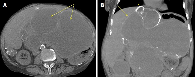
Figure 3 Computed tomography of pseudomyxoma peritonei[26].
This is an axial computed tomography scan. A: Cystic accumulations of mucus (arrows) surrounded by calcified rims; B: A coronal reconstruction representing cystic accumulations in the upper abdomen and the liver.
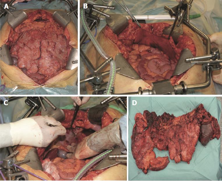
Figure 4 Intraoperative pictures of cytoreductive surgery[11].
This is a figure depicting various stages of cytoreductive surgery. A: A view of a pseudomyxoma patient’s abdominal cavity immediately after laparotomy; B: Stripping of the right anterior peritoneum; C: Depicts stripping of the right subphrenic peritoneum; D: An image of the resected terminal ileum, colon, and spleen affected by pseudomyxoma peritonei.
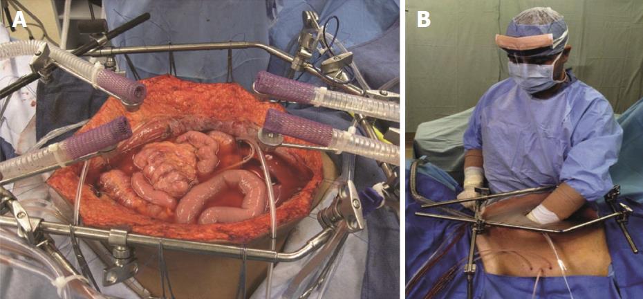
Figure 5 Hyperthermic intraperitoneal chemotherapy after complete cytoreductive surgery[27].
This figure demonstrates the hyperthermic intraperitoneal chemotherapy (HIPEC) setup after cytoreductive surgery has been completed. A: HIPEC administered using an open technique; B: After placement of tubes, drains and temperature probes, the skin edges are elevated onto the rim of the self-retaining retractor using a running suture. A plastic sheet covers the abdomen to prevent splashing and loss of chemotherapy agent. A slit in the sheet allows the surgeon to access the abdominal cavity. Continuous mixing by the surgeon ensures all abdominal surfaces are uniformly coated with doses of heated chemotherapy.
- Citation: Rizvi SA, Syed W, Shergill R. Approach to pseudomyxoma peritonei. World Journal of Gastrointestinal Surgery 2018; 10(5): 49-56
- URL: https://www.wjgnet.com/1948-9366/full/v10/i5/49.htm
- DOI: https://dx.doi.org/10.4240/wjgs.v10.i5.49


