Copyright
©The Author(s) 2017.
World J Gastrointest Oncol. Dec 15, 2017; 9(12): 497-501
Published online Dec 15, 2017. doi: 10.4251/wjgo.v9.i12.497
Published online Dec 15, 2017. doi: 10.4251/wjgo.v9.i12.497
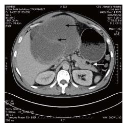
Figure 1 An abdominal computed tomography scan exhibited solid and mixed cystic lesions, measuring > 28 cm diameter (black arrow).
The tumor was apart from the pancreas (white arrow).
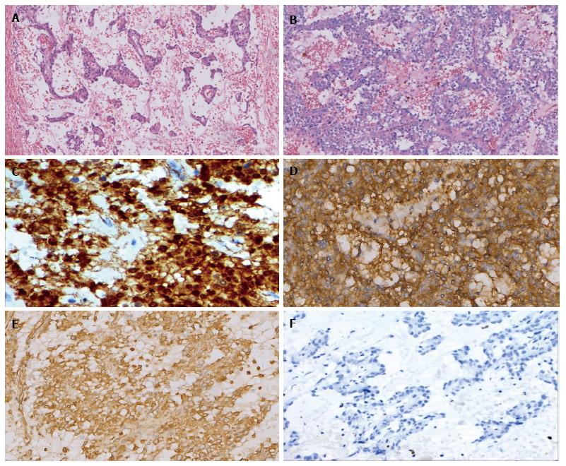
Figure 2 Histological and immunohistochemical findings of the tumor (× 200).
The tumor cells are arranged in solid sheets, pseudopapillary and microcysts (A and B: Hematoxylin-eosin stain), and are immunohistochemically positive for alpha-1-antitrypsin (C), β-catenin (D: Cytoplasmic and nuclear staining), CD56 (E), whereas negative for chromogranin (F).
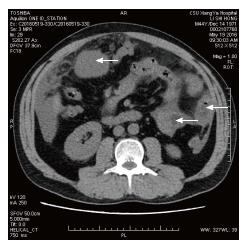
Figure 3 An abdominal computed tomography scan exhibited multiple tumors in peritoneum, greater omentum, and colonic wall (white arrow).
- Citation: Wu H, Huang YF, Liu XH, Xu MH. Extrapancreatic solid pseudopapillary neoplasm followed by multiple metastases: Case report. World J Gastrointest Oncol 2017; 9(12): 497-501
- URL: https://www.wjgnet.com/1948-5204/full/v9/i12/497.htm
- DOI: https://dx.doi.org/10.4251/wjgo.v9.i12.497


