Copyright
©The Author(s) 2016.
World J Gastrointest Oncol. Mar 15, 2016; 8(3): 326-329
Published online Mar 15, 2016. doi: 10.4251/wjgo.v8.i3.326
Published online Mar 15, 2016. doi: 10.4251/wjgo.v8.i3.326
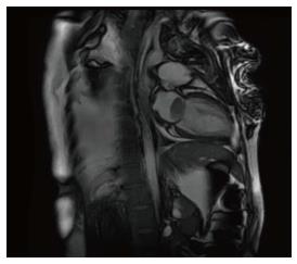
Figure 1 Cardiac magnetic resonance imaging showing a large mass in the left atrium.
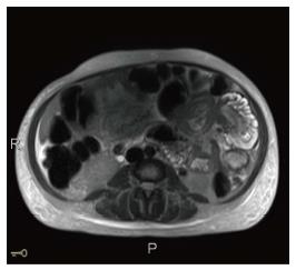
Figure 2 Magnetic resonance imaging of abdomen showing entero-enteric intussusception in axial cut.
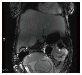
Figure 3 Magnetic resonance imaging of the abdomen showing an entero-enteric intussusception in the left upper quadrant.
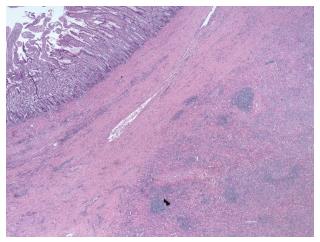
Figure 4 Normal small bowel mucosa (left hand corner) in contrast to infiltrative area of increased cellularity (right lower hand corner).
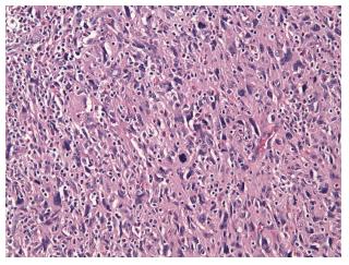
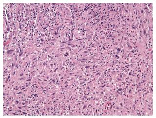
Figure 5 High power: Population of poorly differentiated malignant cells that are high grade (pleomorphic, hyperchromatic and contain increased mitotic activity) and are similar to the intracardiac dedifferentiated liposarcoma.
Figure 6 Poorly differentiated malignant neoplasm composed of variably spindled polygonal or histiocytoid cells and irregular vesicular nuclei consistent with a dedifferentiated liposarcoma.
- Citation: Gomez G, Bilal M, Klepchick P, Clarke K. Rare case of entero-enteric intussusception caused by small bowel metastasis from a cardiac liposarcoma. World J Gastrointest Oncol 2016; 8(3): 326-329
- URL: https://www.wjgnet.com/1948-5204/full/v8/i3/326.htm
- DOI: https://dx.doi.org/10.4251/wjgo.v8.i3.326


