Copyright
©The Author(s) 2016.
World J Gastrointest Oncol. Dec 15, 2016; 8(12): 805-809
Published online Dec 15, 2016. doi: 10.4251/wjgo.v8.i12.805
Published online Dec 15, 2016. doi: 10.4251/wjgo.v8.i12.805
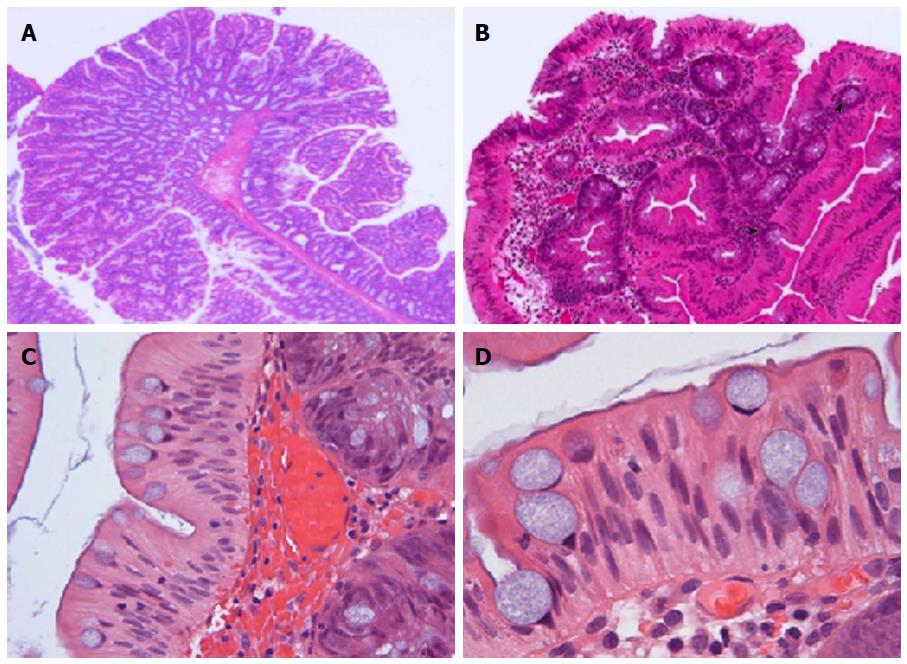
Figure 1 Traditional serrated adenoma.
A: Traditional serrated adenoma (TSA) demonstrating an arborizing, “pinecone-like” pattern; × 12.5; B: TSA replete with abortive crypts or ectopic crypt foci (ECF) (arrowhead); × 100; C: Characteristic slit-like luminal serrations with deep clefts and indentations, resulting in mushroom-like or jigsaw puzzle-like appearance, which resemble the apical brush border of small bowel; × 200; D: The epithelial cells have intensely eosinophilic cytoplasm with centrally located palisaded, regular, pencillate nuclei and nuclear grooves. In addition, there are haphazardly distributed goblet cells with apical mucin and basally located nuclei; × 400 (all H and E).
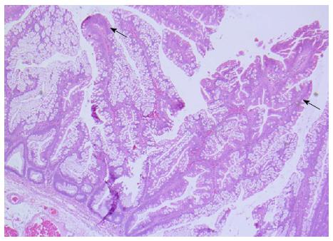
Figure 2 Sessile serrated adenoma with the characteristic dilated crypts.
It is a horizontal growth along the muscularis mucosa and deep serration, seamlessly merging with foci of traditional serrated adenomas like areas (arrow); × 100, H and E.
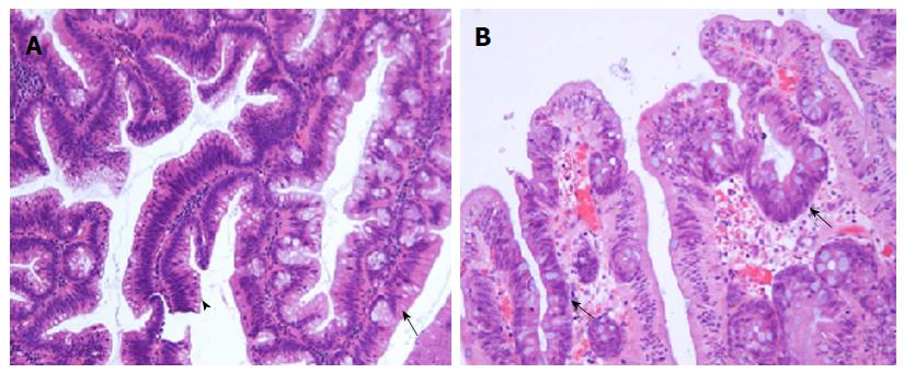
Figure 3 Traditional serrated adenoma and dysplasia.
A: Traditional serrated adenoma (TSA) (arrow), sharply demarcated form more a more conventional tubulovillous adenoma (TVA) (arrowhead); × 100; B: TSA with conventional dysplasia with some preservation of the characteristic architecture; × 200 (both H and E).
- Citation: Kalimuthu SN, Chelliah A, Chetty R. From traditional serrated adenoma to tubulovillous adenoma and beyond. World J Gastrointest Oncol 2016; 8(12): 805-809
- URL: https://www.wjgnet.com/1948-5204/full/v8/i12/805.htm
- DOI: https://dx.doi.org/10.4251/wjgo.v8.i12.805


