Copyright
©2014 Baishideng Publishing Group Inc.
World J Gastrointest Oncol. Sep 15, 2014; 6(9): 311-324
Published online Sep 15, 2014. doi: 10.4251/wjgo.v6.i9.311
Published online Sep 15, 2014. doi: 10.4251/wjgo.v6.i9.311
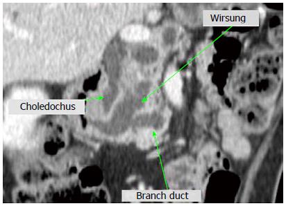
Figure 1 Computerized tomography scan demonstrating massive dilatation of the main pancreatic duct and its branches.
Obstructed bile duct is also dilated.
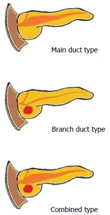
Figure 2 Scheme of macroscopic classification of intraductal papillary mucinous neoplasm.
Figure 3 Macroscopic classification of intraductal papillary mucinous neoplasm.
A: Main duct type; B: Branch duct type; C: Combined type.
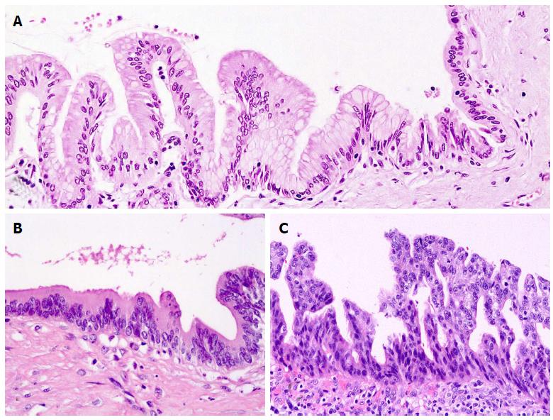
Figure 4 Different degrees of dysplasia.
A: Low-grade (gastric IPMN). See transition with non dysplastic normal duct epithelium (right side); B: Intermediate-grade (intestinal IPMN); C: High-grade (pancreatobiliary IPMN). IPMN: Intraductal papillary mucinous neoplasm.
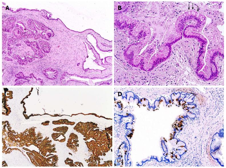
Figure 5 Gastric intraductal papillary mucinous neoplasm.
A: The neoplasm involves branch ducts with a multicystic appearance; B: Columnar cells with basal nucleus and apical mucin. Notice the scattered goblet cells (arrows); C: Immunohistochemical MUC5AC expression (colored brown by diaminobenzidine); D: MUC2 highlighting the goblet cells. MUC: Mucin.
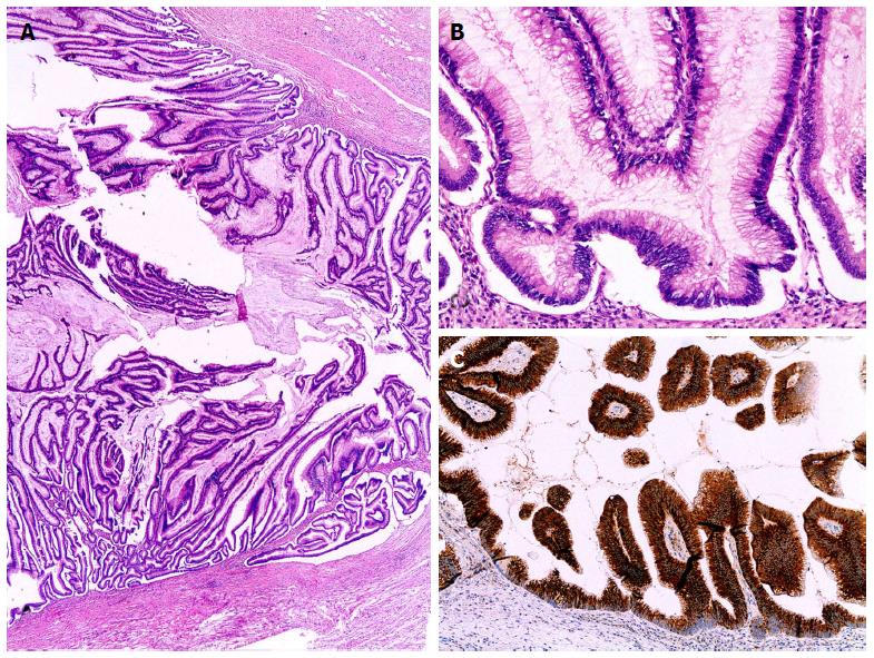
Figure 6 Intestinal intraductal papillary mucinous neoplasm.
A: Main duct distended by long papillae; B: Projections of columnar cells with pseudostratified nuclei; C: Immunohistochemical MUC2 expression. MUC: Mucin.
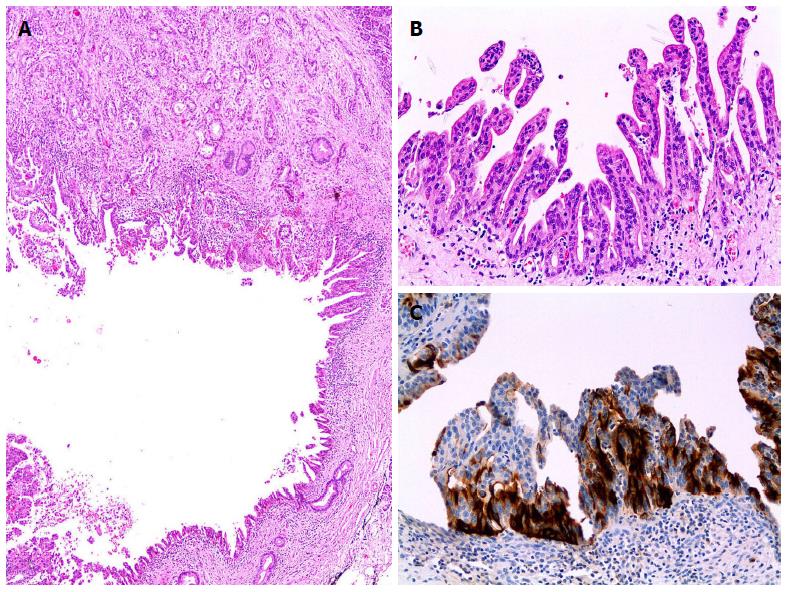
Figure 7 Pancreatobiliary intraductal papillary mucinous neoplasm.
A: Intraductal papillary mucinous neoplasm with associated conventional duct carcinoma (upper side); B: Small thin papillae with cuboidal neoplastic epithelium; C: Immunohistochemical MUC1 expression. MUC: Mucin.
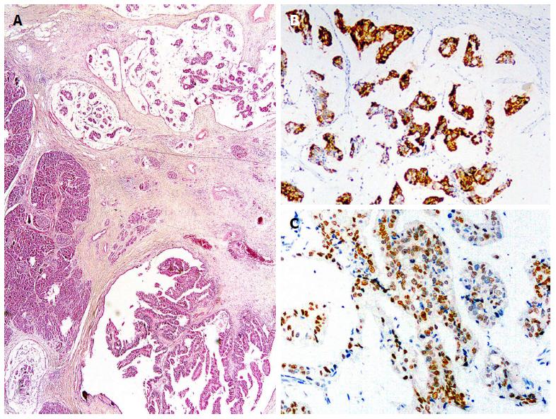
Figure 8 Colloid carcinoma.
A: Invasive neoplastic cells floating in pools of mucin (upper side) and associated with intraductal papillary mucinous neoplasm (lower side); B: Immunohistochemical MUC2 expression; C: CDX2 nuclear immunoexpression. MUC: Mucin.
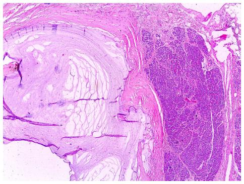
Figure 9 Pseudoinvasion.
Mucin spillage dissecting into the pancreatic stroma without neoplastic cells.
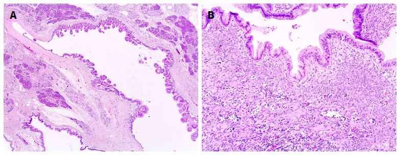
Figure 10 Mucinous cystic neoplasm.
A: An example with papillary projections and surrounded by a thick collagenized band; B: Demonstration of ovarian-type stroma, at least focally, leads to diagnosis.
- Citation: Castellano-Megías VM, Andrés CID, López-Alonso G, Colina-Ruizdelgado F. Pathological features and diagnosis of intraductal papillary mucinous neoplasm of the pancreas. World J Gastrointest Oncol 2014; 6(9): 311-324
- URL: https://www.wjgnet.com/1948-5204/full/v6/i9/311.htm
- DOI: https://dx.doi.org/10.4251/wjgo.v6.i9.311


