Copyright
©2014 Baishideng Publishing Group Co.
World J Gastrointest Oncol. Feb 15, 2014; 6(2): 52-54
Published online Feb 15, 2014. doi: 10.4251/wjgo.v6.i2.52
Published online Feb 15, 2014. doi: 10.4251/wjgo.v6.i2.52
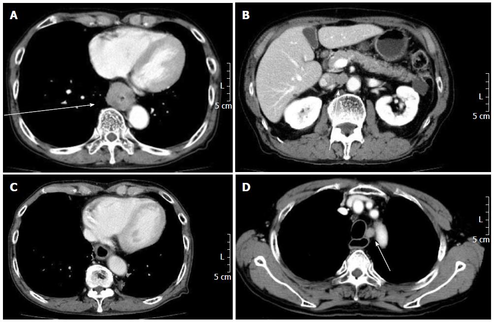
Figure 1 Computed tomography.
A: Circumferential wall thickening and a 39 mm × 35 mm tumor on the lower thoracic esophagus; B: Swelling of the abdominal para-aortic lymph node; C: A remarkable shrinking of the mass in the lower esophagus; D: Swelling of the left para-tracheal lymph node.
- Citation: Yamashita H, Okuma K, Nomoto A, Yamashita M, Igaki H, Nakagawa K. Extended cancer-free survival after palliative chemoradiation for metastatic esophageal cancer. World J Gastrointest Oncol 2014; 6(2): 52-54
- URL: https://www.wjgnet.com/1948-5204/full/v6/i2/52.htm
- DOI: https://dx.doi.org/10.4251/wjgo.v6.i2.52


