Copyright
©2012 Baishideng.
World J Gastrointest Oncol. Jul 15, 2012; 4(7): 184-186
Published online Jul 15, 2012. doi: 10.4251/wjgo.v4.i7.184
Published online Jul 15, 2012. doi: 10.4251/wjgo.v4.i7.184
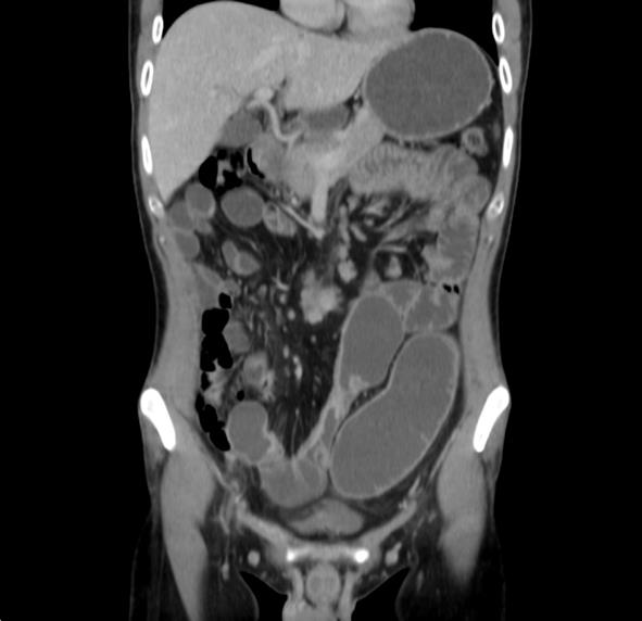
Figure 1 Abdominal computed tomography scan revealing a classic radiological finding of Crohn’s disease: Thickened jejunal wall with pre stenotic dilatation, as well as congestion of the mesenterial blood vessels.
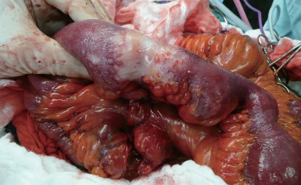
Figure 2 Primary adenocarcinoma of the jejunum, after dividing the mesenterial vascular supply.
- Citation: Drukker L, Edden Y, Reissman P. Adenocarcinoma of the small bowel in a patient with occlusive Crohn’s disease. World J Gastrointest Oncol 2012; 4(7): 184-186
- URL: https://www.wjgnet.com/1948-5204/full/v4/i7/184.htm
- DOI: https://dx.doi.org/10.4251/wjgo.v4.i7.184


