Copyright
©2012 Baishideng Publishing Group Co.
World J Gastrointest Oncol. Dec 15, 2012; 4(12): 250-258
Published online Dec 15, 2012. doi: 10.4251/wjgo.v4.i12.250
Published online Dec 15, 2012. doi: 10.4251/wjgo.v4.i12.250
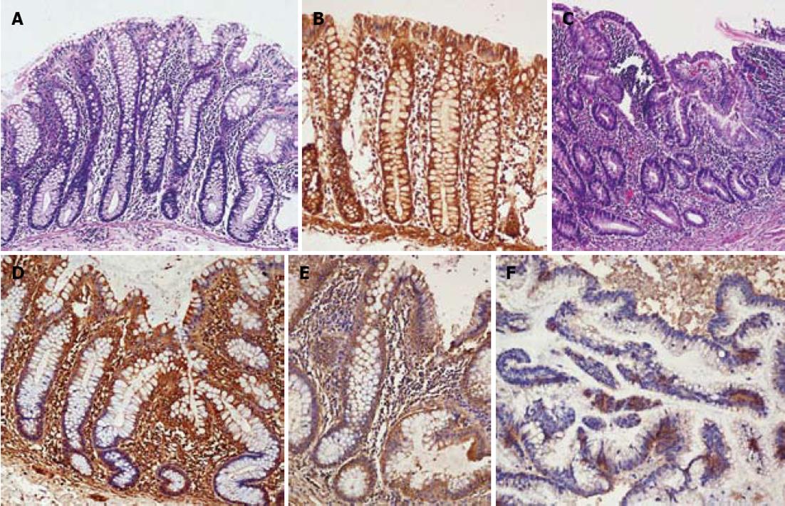
Figure 1 Medium power photomicrograph.
A: Medium power photomicrograph of colonic mucosa taken from the areas of granular mucosa showing hyperplastic aberrant crypt focus (ACF) [hematoxylin eosin (HE), × 300]; B: Medium power photomicrograph of normal colonic mucosa showing diffuse and strong expression for Fhit protein [peroxidase anti-peroxidase (PAP), × 300]; C: Medium power photomicrograph of colonic mucosa taken from the granular mucosa closed to the tumor showing dysplastic ACF (HE, × 250); D: Medium power photomicrograph of hyperplastic ACF showing diffuse but weaker cytoplasmic Fhit protein expression (PAP, × 300); E: Medium power photomicrograph of dysplastic ACF showing weak cytoplasmic Fhit protein expression (PAP, × 250); F: Medium power photomicrograph of a colonic adenocarcinoma showing a negative staining for Fhit protein (PAP, × 250).
- Citation: Vaiphei K, Rangan A, Singh R. Aberrant crypt focus and fragile histidine triad protein in sporadic colorectal carcinoma. World J Gastrointest Oncol 2012; 4(12): 250-258
- URL: https://www.wjgnet.com/1948-5204/full/v4/i12/250.htm
- DOI: https://dx.doi.org/10.4251/wjgo.v4.i12.250


