Copyright
©2011 Baishideng Publishing Group Co.
World J Gastrointest Oncol. Jun 15, 2011; 3(6): 103-106
Published online Jun 15, 2011. doi: 10.4251/wjgo.v3.i6.103
Published online Jun 15, 2011. doi: 10.4251/wjgo.v3.i6.103
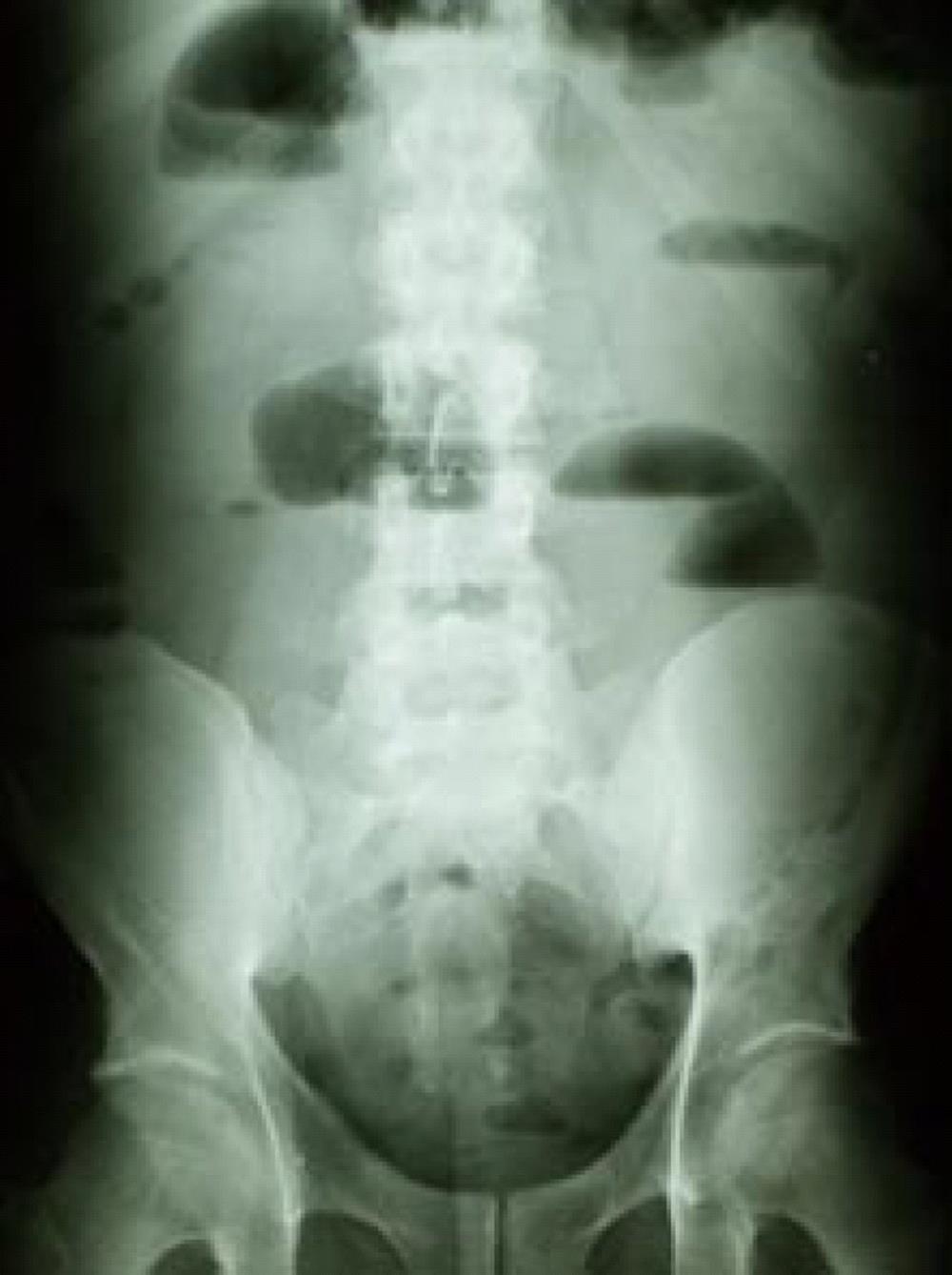
Figure 1 Plain abdominal film showed dilated bowel loops and air-fluid levels.
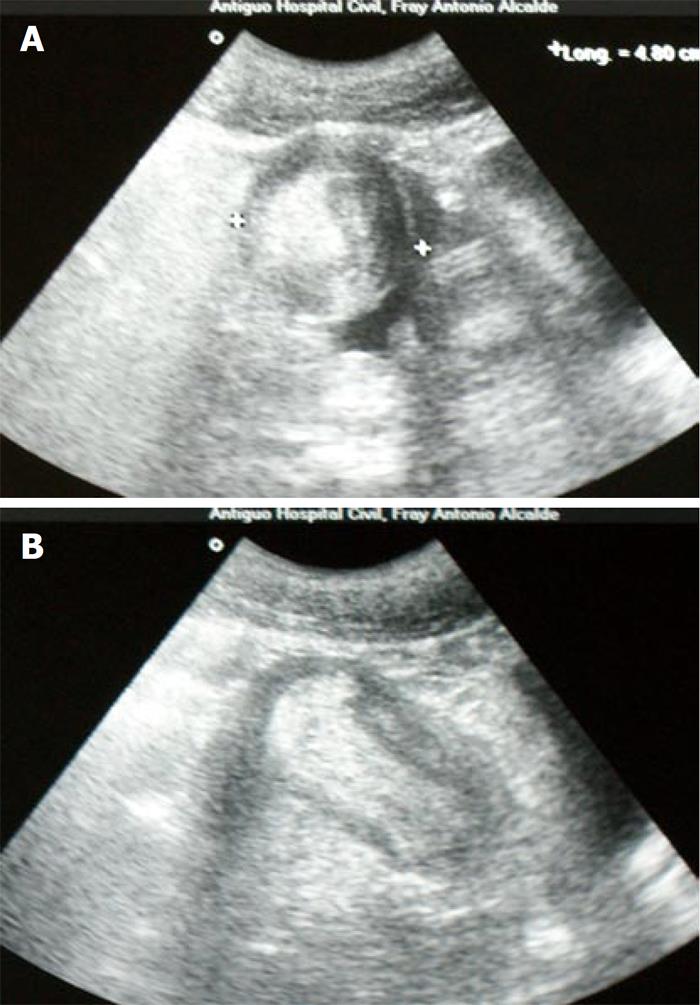
Figure 2 Ultrasonographic feature of a “target” sign on a transverse view (A), and a “sausage-shaped image” in a longitudinal view (B).
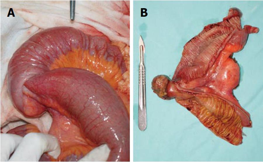
Figure 3 Distended bowel proximal to the intussusception (A), open surgical specimen showing a 3 cm pedunculated-type polypoid tumor (B).
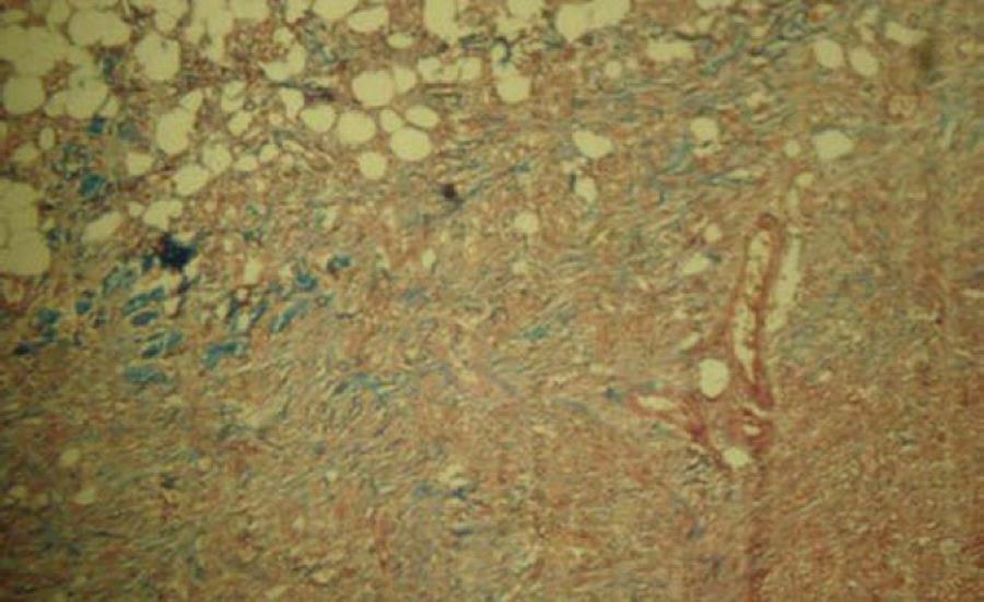
Figure 4 Histopathologic appearance of the ileal tumor showing an active mesenchymal lesion, with no malignant transformation (Masson’s trichrome stain, × 10).
- Citation: Nuño-Guzmán CM, Arróniz-Jáuregui J, Espejo I, Solís-Ugalde J, Gómez-Ontiveros JI, Vargas-Gerónimo A, Valle-González J. Adult intussusception secondary to an ileum hamartoma. World J Gastrointest Oncol 2011; 3(6): 103-106
- URL: https://www.wjgnet.com/1948-5204/full/v3/i6/103.htm
- DOI: https://dx.doi.org/10.4251/wjgo.v3.i6.103


