Copyright
©2010 Baishideng Publishing Group Co.
World J Gastrointest Oncol. Dec 15, 2010; 2(12): 446-448
Published online Dec 15, 2010. doi: 10.4251/wjgo.v2.i12.446
Published online Dec 15, 2010. doi: 10.4251/wjgo.v2.i12.446
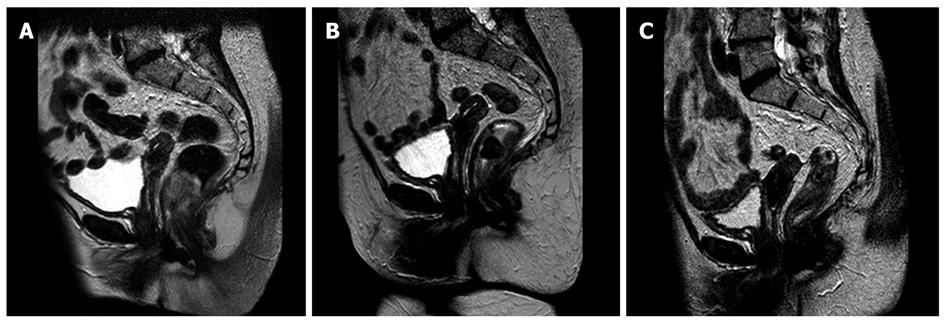
Figure 1 Magnetic resonance imaging images of the patient.
A: Initial magnetic resonance imaging (MRI) scan showing rectal wall invasion to over one third of the circumference of the rectum, extending beyond the rectal wall; B: MRI scan performed 2 mo following completion of treatment: a favourable response was reported with a reduction in size from 30 to 19 mm, but with a remaining suspicious area of rectal wall extension (T3); C: MRI scan 4 mo post excision showed no residual disease.
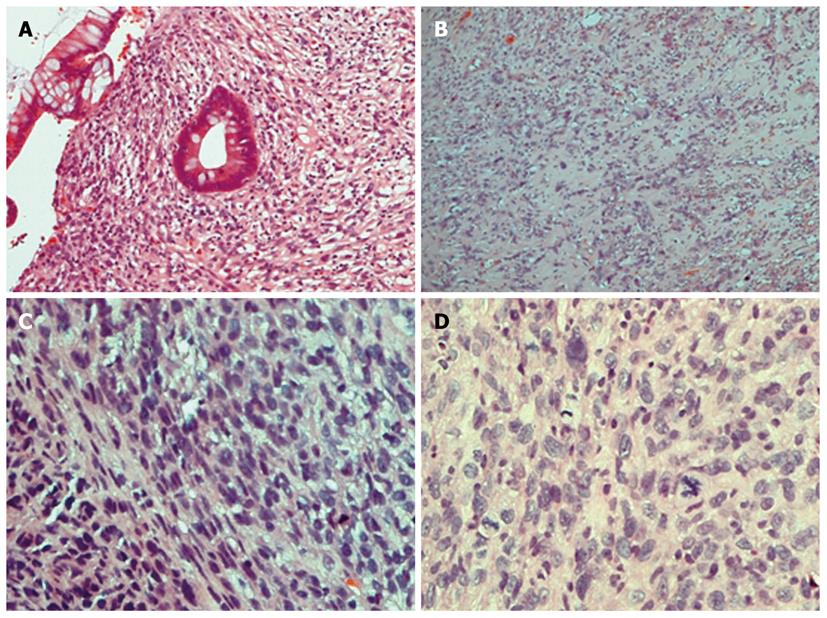
Figure 2 Hematoxylin and eosin stain slides of initial anal biopsy pathology specimen.
A: Anal mucosa showing infiltration by tumour; B: Pleomorphic cytology and fibrotic changes in some areas of tumour; C: Tumour with spindle and epithelioid nuclei; D: Tumour with pleomorphism and frequ.
- Citation: Mikropoulos C, Williams T, Munthali L, Summers J. A rare case of anal tumor: Anal carcinosarcoma. World J Gastrointest Oncol 2010; 2(12): 446-448
- URL: https://www.wjgnet.com/1948-5204/full/v2/i12/446.htm
- DOI: https://dx.doi.org/10.4251/wjgo.v2.i12.446


