Copyright
©2010 Baishideng Publishing Group Co.
World J Gastrointest Oncol. Oct 15, 2010; 2(10): 395-398
Published online Oct 15, 2010. doi: 10.4251/wjgo.v2.i10.395
Published online Oct 15, 2010. doi: 10.4251/wjgo.v2.i10.395
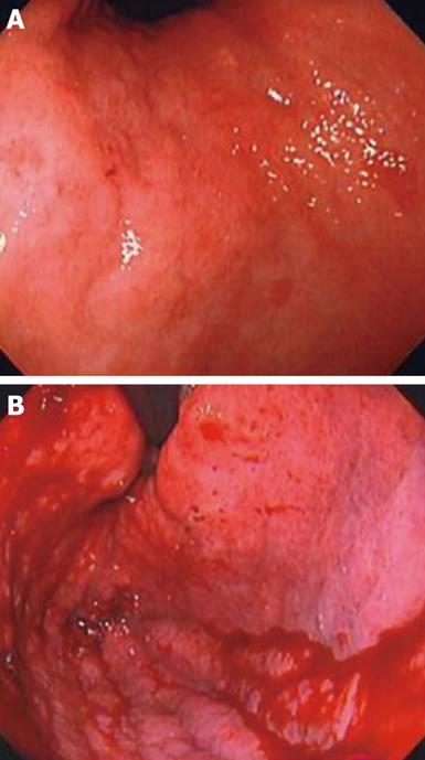
Figure 1 Gastrointestinal endoscopy on admission showed Type 4 tumor spread from the gastric upper third of the stomach and body, with loss of distensibility of the gastric wall.
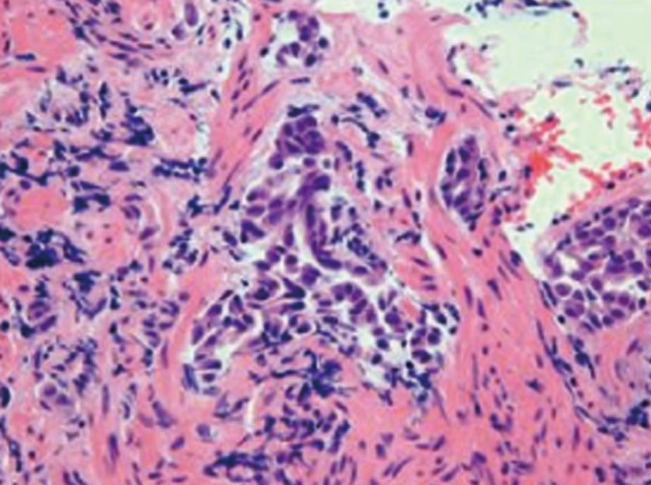
Figure 2 Hematoxylin and eosin staining of the specimen from the gastric tumor showed adenocarcinoma cells infiltrated in the gastric mucosa.
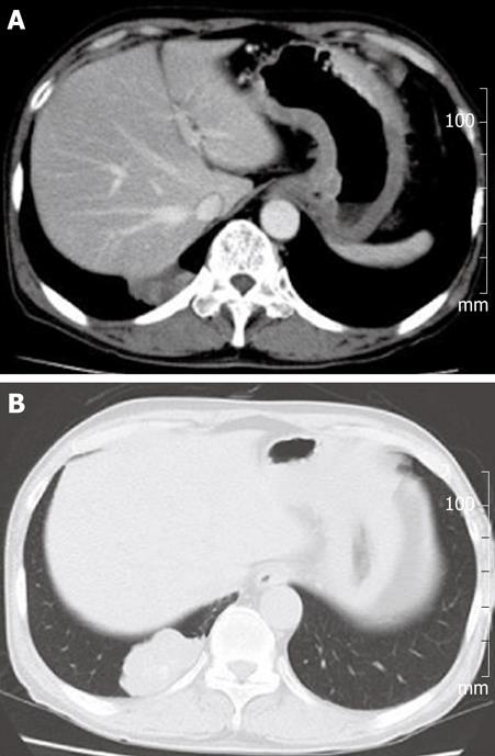
Figure 3 Chest CT on admission showed thickening of the gastric wall (A) and mass shadow in the right lung S10 segment (A and B).
Figure 4 Immunohistochemical staining of gastric cancer for Cytokeratin 7 (A) and TTF-1 (B) showed immunopositive staining for both markers.
- Citation: Okazaki R, Ohtani H, Takeda K, Sumikawa T, Yamasaki A, Matsumoto S, Shimizu E. Gastric metastasis by primary lung adenocarcinoma. World J Gastrointest Oncol 2010; 2(10): 395-398
- URL: https://www.wjgnet.com/1948-5204/full/v2/i10/395.htm
- DOI: https://dx.doi.org/10.4251/wjgo.v2.i10.395


