
















Copyright: ©Author(s) 2026.
World J Gastrointest Oncol. Nov 15, 2026; 18(11): 124384
Published online Nov 15, 2026. doi: 10.4251/wjgo.124384
Published online Nov 15, 2026. doi: 10.4251/wjgo.124384
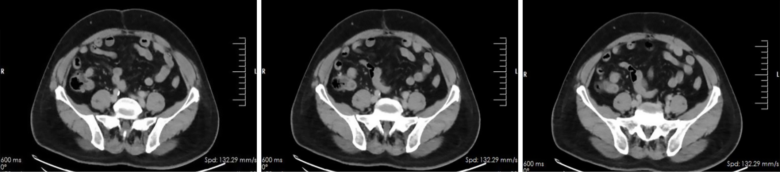
Figure 1 Contrast-enhanced abdominal computed tomography findings.
Computed tomography scan showing an elongated and thickened appendix without obvious lymphadenopathy or distant metastases.
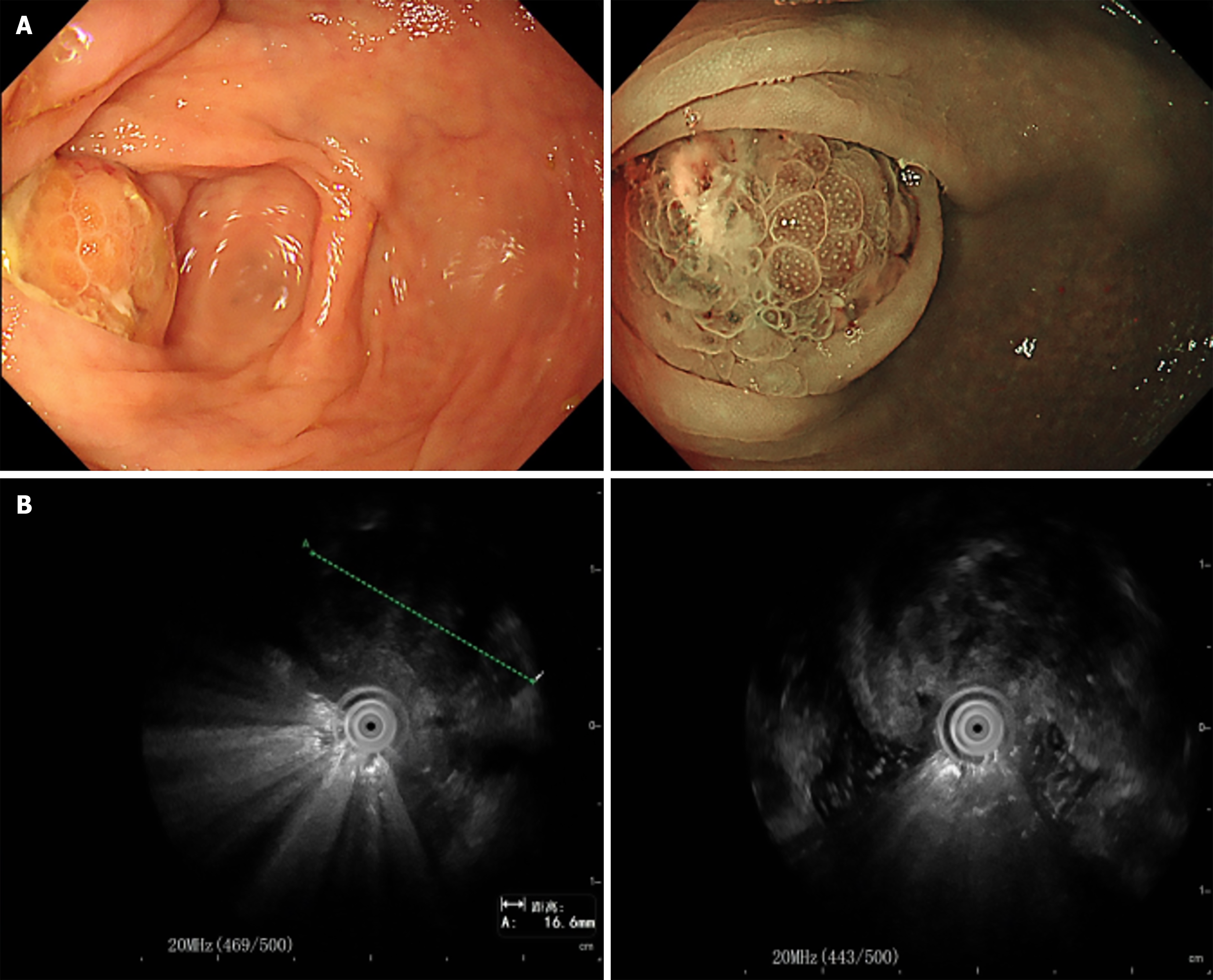
Figure 2 Endoscopic ultrasound showing a mixed hypoechoic lesion at the appendiceal orifice.
A: White-light endoscopy; B: Endoscopic ultrasound image.
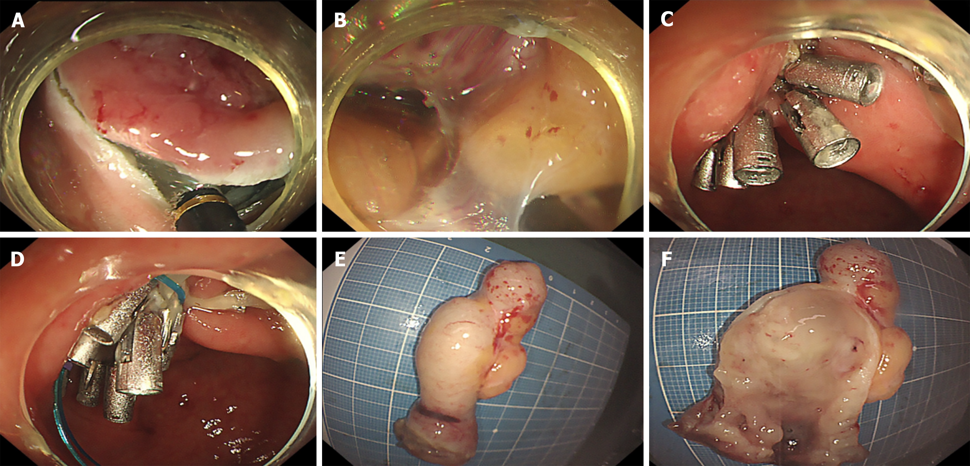
Figure 3 Intraoperative endoscopic images.
A and B: Full-thickness resection; C and D: Closure of defect with metal clips; E and F: Excised specimen.
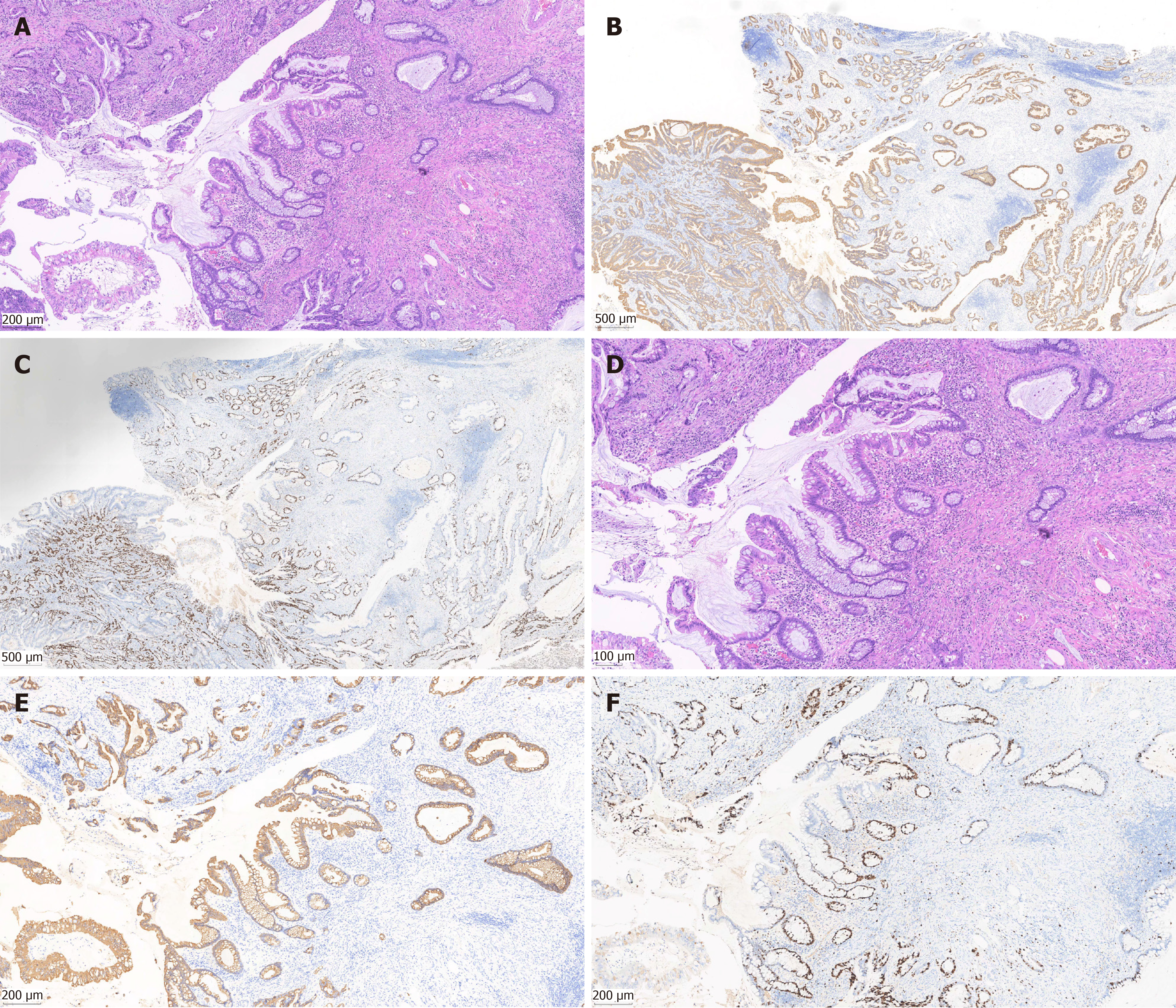
Figure 4 Histopathology and immunohistochemistry.
A: Hematoxylin and eosin stain showing crypt branching and serrated morphology (× 40); B: Diffuse cytokeratin 20 positivity (× 40); C: Asymmetric Ki-67 expression (× 40); D: Hematoxylin and eosin stain showing crypt branching and serrated morphology (× 100); E: Diffuse cytokeratin 20 positivity (× 100); F: Asymmetric Ki-67 expression (× 100).
- Citation: Liu YL, Jiang WJ, Xu JQ, Huang LL, Liu J, Wang YT. Appendiceal adenocarcinoma arising from serrated lesions: A case report. World J Gastrointest Oncol 2026; 18(11): 124384
- URL: https://www.wjgnet.com/1948-5204/full/v18/i11/124384.htm
- DOI: https://dx.doi.org/10.4251/wjgo.124384