
















Copyright: ©Author(s) 2026.
World J Gastrointest Oncol. Nov 15, 2026; 18(11): 121634
Published online Nov 15, 2026. doi: 10.4251/wjgo.121634
Published online Nov 15, 2026. doi: 10.4251/wjgo.121634
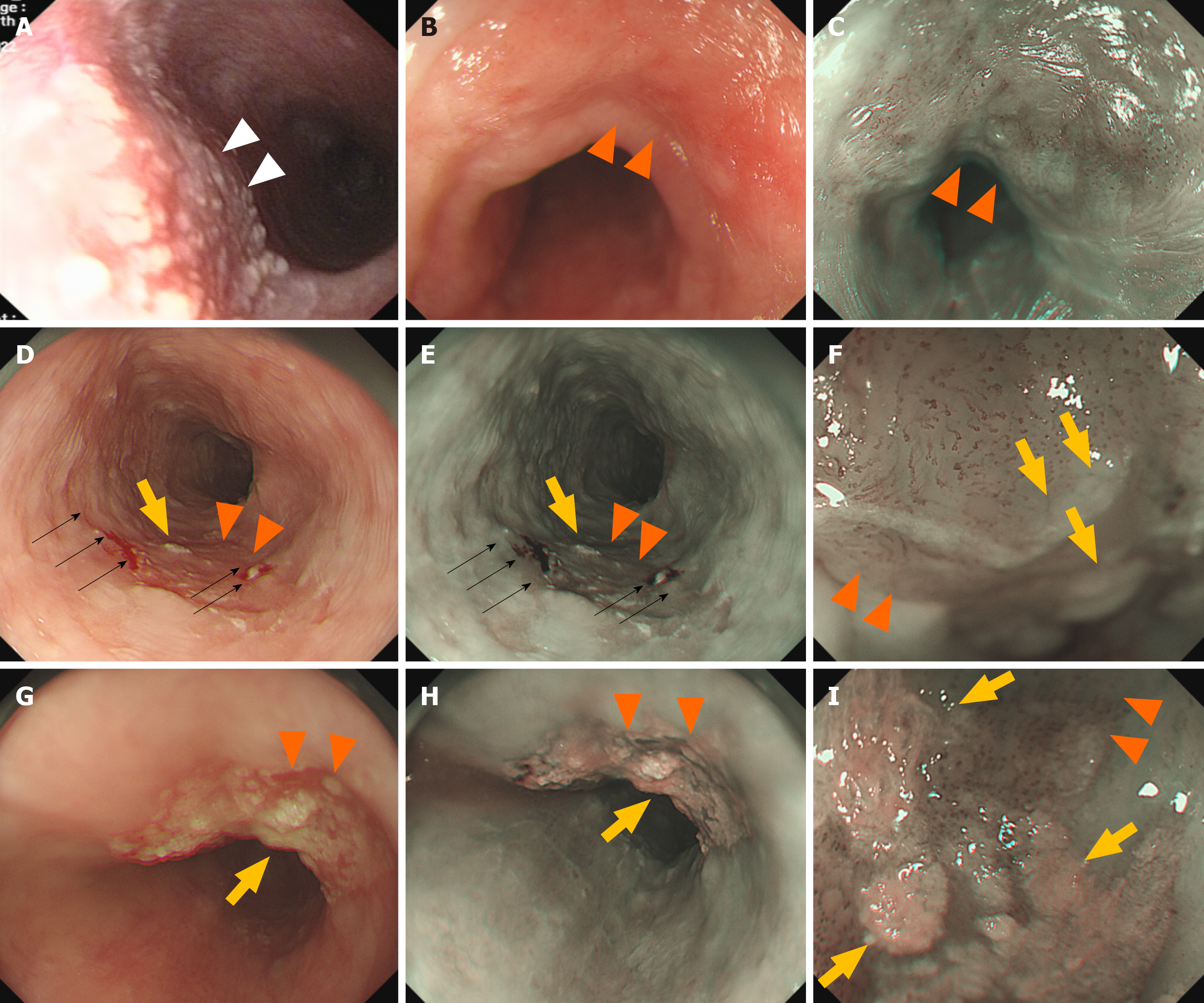
Figure 1 Endoscopic appearance of early esophageal squamous cell carcinomas and esophageal keratosis.
A: Esophageal keratosis (white arrows) on the surface of normal esophageal epithelium under white light endoscopy (WLE); B: Nonepithelial keratosis in early esophageal squamous cell carcinoma (ESCC) (orange arrows) under WLE; C: Nonepithelial keratosis early ESCC (orange arrows) under narrow-band imaging with magnifying endoscopy (NBI-ME) (orange arrows); D: Proportion of esophageal keratinized (yellow arrows) area < 30% in early ESCC (orange arrows) under WLE; spontaneous bleeding was visible (black arrows) in the lesion area; E: Proportion of esophageal keratinized (yellow arrows) area < 30% in early ESCC (orange arrows) under low-power NBI-ME; spontaneous bleeding was visible (black arrows) in the lesion area; F: Proportion of esophageal keratinized (yellow arrows) area < 30% in early ESCC (orange arrows) under high-power NBI-ME; spontaneous bleeding was visible (black arrows) in the lesion area; G: Proportion of esophageal keratinized (yellow arrows) area ≥ 30% in early ESCC (orange arrows) under WLE; H: Proportion of esophageal keratinized (yellow arrows) area ≥ 30% in early ESCC (orange arrows) under low-power NBI-ME; I: Proportion of esophageal keratinized (yellow arrows) area ≥ 30% in early ESCC (orange arrows) under high-power NBI-ME.
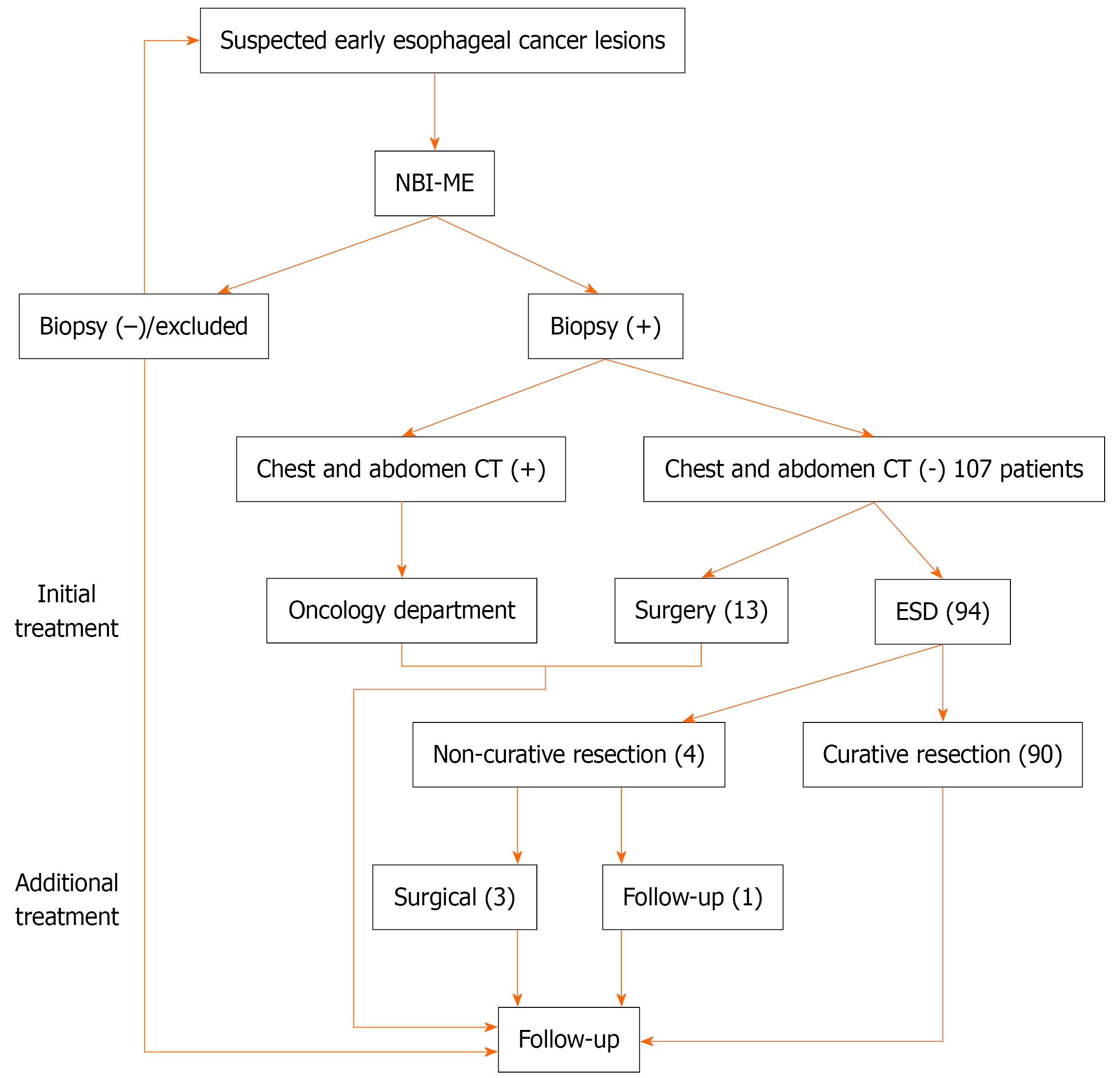
Figure 2 Flowchart for the management of suspected early esophageal squamous cell carcinoma.
NBI-ME: Narrow band imaging with magnifying endoscopy; CT: Computed tomography; ESD: Endoscopic mucosal dissection.
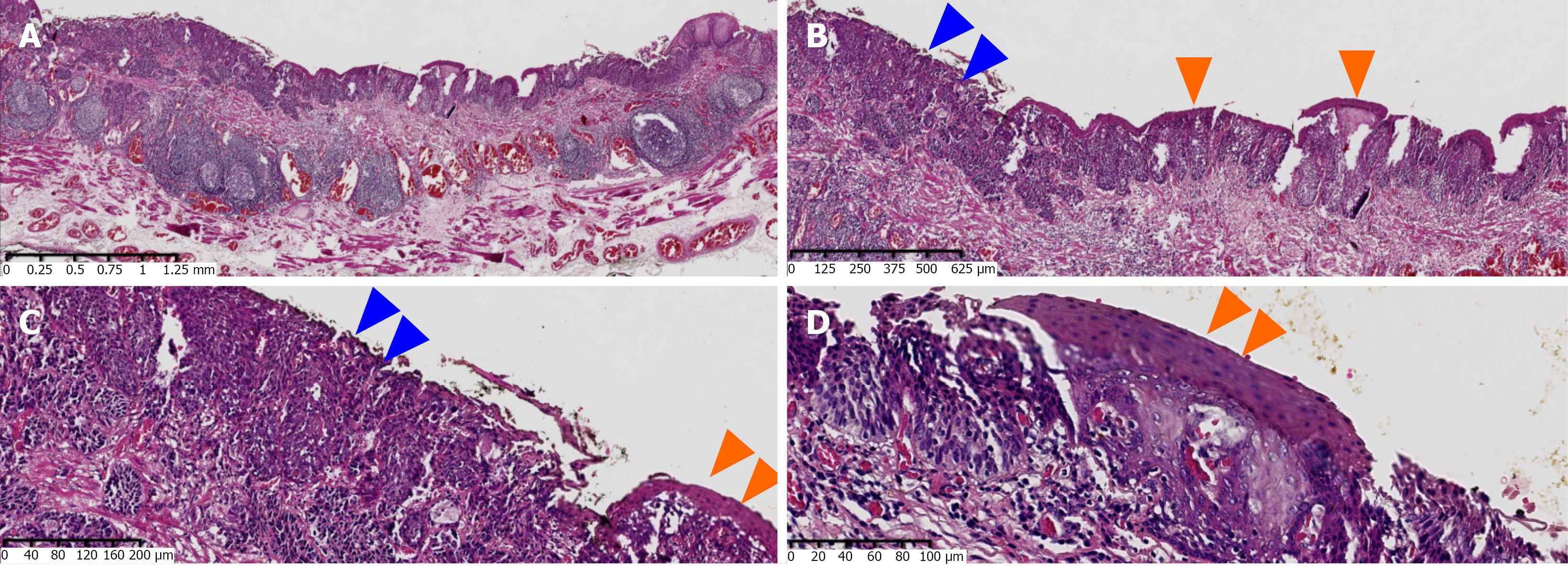
Figure 3 Hematoxylin and eosin staining of resected specimens obtained through endoscopic mucosal dissection of epithelial keratosis in early Esophageal squamous cell carcinoma.
A: 10 ×; B: 40 ×; C: 100 ×; D: 200 ×. Blue arrows indicate tumor areas not covered by epithelial keratosis; orange arrows indicate tumor surfaces covered with esophageal keratinization.
- Citation: Zhang YJ, Wen W, Li F, Yuan J, Zou DH, Shi HP, Liang C, Yang Y. Proportion of esophageal keratinized area ≥ 30% reduces accuracy of preoperative endoscopic evaluation in early esophageal squamous cell carcinoma. World J Gastrointest Oncol 2026; 18(11): 121634
- URL: https://www.wjgnet.com/1948-5204/full/v18/i11/121634.htm
- DOI: https://dx.doi.org/10.4251/wjgo.121634